Understanding Human Anatomy: A Comprehensive Guide to Anatomical Directions and Tissues
280 likes | 393 Views
Dive into the intricate world of human anatomy and physiology with this insightful overview. Learn about anatomical directions, including terms like anterior, posterior, medial, and lateral. Explore various tissue types—epithelial, connective, muscle, and nervous—and their specific characteristics and functions. Understand the structural roles of epithelial tissues and the properties of connective tissue, including cartilage and bone. Finally, familiarize yourself with muscle tissue types and their vital roles in movement and body functions.

Understanding Human Anatomy: A Comprehensive Guide to Anatomical Directions and Tissues
E N D
Presentation Transcript
Human Anatomy & Physiology Mrs. Hodges Room A204 Per 1, 2, 3
Anatomical Directions • Anatomical position • Illustrated at the left • Anatomical Directions-(for the biped) • Anterior (ventral) vs. Posterior (dorsal) • Medial vs. Lateral • Superior (cranial) vs. Inferior (caudal) • Superficial vs. Deep • Proximal vs. Distal • Anatomical Planes • Frontal = Coronal • Transverse = Cross Section • Sagittal
Cell Connections • Cells are connected to neighboring cells via: • Proteins – adjacent proteins in membranes fuse to form: • Cell Junctions • Tight Junctions - plasma membrane of adjacent cells fuse; impermeable • Desmosomes-adhesive spots on lateral sides • Gap junction-spot-like junction occurring anywhere, lets small molecules pass
Histology Study of tissues A tissue is a group of cells with similar structure and embryonic origin working together to perform a particular function in the body.
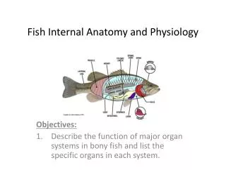
Tissues: groups of cells closely associated that have a similar structure and perform a related function • Four types of tissue A. Epithelial = covering/lining B. Connective = support C. Muscle = movement D. Nervous = control • Most organs contain all 4 types
A. EPITHELIAL TISSUE: sheets of cells that cover a surface or line a cavity • Functions • Protection • Secretion • Absorption
How are epithelial tissues classified? • Shape • Squamous • Cuboidal • Columnar • Number of Layers • Simple: single layer • Stratified: many layers
8 Specific Epithelial Tissues Simple • Simple squamous • Simple cuboidal • Simple columnar • Pseudostratified
8 Specific Epithelial Tissues Simple • Simple squamous • Simple cuboidal • Simple columnar • Pseudostratified Stratified • Stratified squamous • Stratified cuboidal • Stratified columnar • transitional
Quiz!! Can You Identify the Classes of Epithelium? E D A B C
Structural Characteristics of Epithelium • Cellularity • Mostly composed of cell • Specialized Contacts • Composed mostly of sheets • Polarity • Has one free surface, the other is attached to an underlying tissue • Avascular • No blood vessels • Regenerative • Replaces cells with like cells • Basement Membrane • Is the foundation
B. CONNECTIVE TISSUE Structural Characteristics • Cells • Fibro- -blast = immature cell that secretes matrix • Hemocyto- • Chondro- -cyte = mature cell that maintains matrix • Osteo- • Extracellular matrix Tissue component that is NOT the cells and is made up of: • ground substance = amorphous substance that fills space between cells and consists of interstitial fluid, proteins and polysaccharides. The more polysaccharides the stiffer the ground substance. • fibers = interspersed throughout the ground substance and provides strength to the matrix.
FIBER TYPES • Collagen (aka white) – • Tough • stronger than steel fibers of same size • provide high tensile strength (resists longitudinal stress). • Elastic (aka yellow) – • Can be stretched to 1.5X its length • recoil to original size • found where great elasticity is needed • Reticular – • Fine collagenous fibers that form a delicate branching network within solid organs such as spleen and liver.
4 Types of Connective Tissue 1. Connective Tissue Proper Made by fibroblasts 2. Cartilage Made by chondroblasts 3. Bone Tissue Made by osteoblasts 4. Blood Made by hemocytoblasts
1) Connective Tissue Proper LOOSE • Areolar • Adipose • Reticular DENSE • Regular • Irregular • Elastic
2) Cartilage • Chondroblastsproduce cartilage tissue • More abundant in embryo than adult • Firm, Flexible • Resists compression • (eg) trachea, meniscus • 80% water • Avascular, NOT Innervated (that means no blood, no pain)
Cartilage in the Body Three types: • Hyaline • most abundant • support via flexibility/resilience • found at limb joints, ribs, nose • very fine collagen fibers • Elastic • many elastic fibers in matrix • great flexibility • Found external ear, epiglottis • Fibrocartilage • resists both compression and tension • found in menisci, intervertebral discs
3) Bone Tissue • Compact • cells contained in spaces called lacuna • fine collagen fibers • ground substance contains minerals • Spongy (Cancellous) • Looks like a sponge • Spaces are filled with red bone marrow which is hematopoietic tissue
4) Blood • Formed by hemocytoblastsin red bone marrow which is hematopoietic tissue • Functions: • Transports waste, gases, nutrients, hormones through cardiovascular system • Helps regulate body temperature • Protects body by fighting infection • Cells • erythrocytes • leukocytes • thrombocytes • Matrix = Plasma
C. MUSCLE TISSUE Consists of cells that are specialized for generating a contraction. Cells are elongated and can become shorter and thicker. Three Types: Skeletal, Cardiac, Smooth
MUSCLE TISSUE FUNCTIONS • Produce movement • Generate heat • Maintain posture • Stabilize joints • Characteristics common to ALL muscle tissue: • made of many cells close together • well vascularized tissue • elongated cells • contain myofilaments ( contractile proteins actin and myosin)
Skeletal Muscle Tissue(each gross skeletal muscle is an organ) • Cells • Long and cylindrical, in bundles • Multinucleate • Obvious Striations • Voluntary • Attached to bones, fascia, skin pg 235
Cardiac Muscle Be Mine • Cells • Found only in the heart • Branching cells • uninucleated • Striations • Connected by Intercalated discs • Cardiac Muscle-Involuntary
Smooth Muscle Tissue • Cells • Single cells, uninucleate • No striations • Involuntary • 2 layers-opposite orientation (circular and longitudinal arrangement) • Found in hollow, muscular organs including blood vessels
D. Nervous Tissue • Neurons: specialized nerve cells • Cell body, dendrite, axon • Brain, spinal cord, nerves