Endocrine System
Endocrine System. The endocrine system is composed of several ductless glands , clusters of cells located within certain organs, & isolated endocrine cells [so-called diffuse neuroendocrine system cells (DNES)] in the epithelial lining of the gastrointestinal & respiratory systems.

Endocrine System
E N D
Presentation Transcript
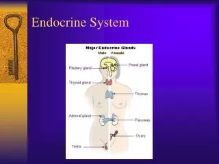
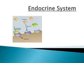
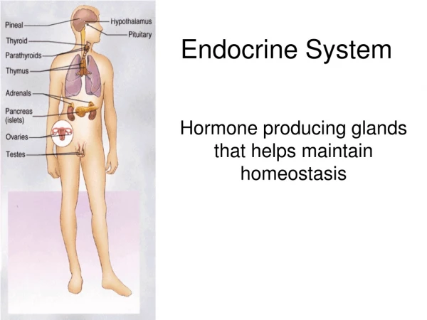
Endocrine System • The endocrine system is composed of several ductless glands, clusters of cells located within certain organs, & isolated endocrine cells [so-called diffuse neuroendocrine system cells (DNES)] in the epithelial lining of the gastrointestinal & respiratory systems. • Glands of the endocrine system include the pituitary, thyroid, parathyroid, adrenal, & pineal glands. • Function. The endocrine system secretes hormones into nearby capillaries & interacts with the nervous system to modulate & control the body’s metabolic activities.
Hormones • Hormones (Gr. hormaein, to excite) are chemical messengers that are carried via the bloodstream to distant target cells. Its functions are essential in maintaining homeostasis and coordinating body growth & development.
Hormones • Steroids, cholesterol-derived compounds • gonadal & adrenocortical steroids • Small peptides, polypeptides, & proteins • insulin, glucagon, GH, ACTH, FSH, LH, ADH, oxytocin, interleukins, various growth factors • Amino acids & arachidonic acid analogs & derivatives • catecholamines, prostaglandins, & leukotriens • thyroid hormones
Hormone’s Receptors • Cell surface receptors • peptide hormones or catecholamines • second messengers • adenylate cyclase/cAMP system • guanyly cyclase/cGMP system • tyrosine kinase system • phosphatidylinositol system • activation of ion channels • Intracellular receptors • steroids & thyroid hormones • influence gene expression directly
Pituitary Gland (Hypophysis) • The pituitary gland lies below the hypothalamus, to which it is structurally & functionally connected (“master organs” of the endocrine system). • It is divided into 2 major subdivisions, the adenohypophysis & the neurohypophysis. • Each subdivision is derived from a distinct embryonic analog, which is reflected in its unique cellular constituents & functions.
Adenohypophysis • The adenohypophysis is also called the anterior pituitary gland. • It is derived from an evagination of the ectoderm of the oropharynx (Rathke’s pouch). • It is subdivided into the pars distalis, pars intermedia, & pars tuberalis.
Pars Distalis • The pars distalis is supported by a connective tissue capsule & framework. • It consists of irregular cords of parenchymal cells lying adjacent to fenestrated sinusoidal capillaries.
Chromophils • Chromophils are parenchymal cells that stain intensely due to their hormone-containing secretory granules. They synthesize, store, & release several hormones. They are regulated by specific stimulatory & inhibitory hormones produced by neurosecretory cells in the hypothalamus & are conveyed to the pars distalis via a system of portal blood vessels originating in the median eminence. • Chromophils are classified into 2 types, depending on the dyes they bind using special histologic stains. With hematoxylin-eosin stain, the distinction between the 2 cell types is much less obvious.
Acidophils • Acidophils bind acid dyes & often stain orange or red. They are small cells of 2 subtypes: somatotropes & lactotropes. • Somatotropes, which produce somatotropin (GH), are stimulated by GHRH or ghrelin & are inhibited by somatostatin. • Lactotropes (mammotropes) produce prolactin, which is stored in small secretory granules. They are stimulated by TRH or VIP & are inhibited by dopamine.
Basophils • Basophils bind basic dyes, typically stain blue & react PAS reagent. They include 3 subtypes: corticotropes, gonadotropes & thyrotropes. • Corticotropes produce proopiomelanocortin (POMC) - ACTH, b-lipotropic hormone (b-LPH), melanocyte-stimulating hormone (MSH), b-endorphin, & enkephalin. They are stimulated by CRH.
Basophils • Gonadotropes produce FSH & LH in both sexes, although in men the latter is sometimes referred to as interstitial cell-stimulating hormone (ICSH). Gonadotropes are stimulated by GnRH. • Thyrotropes produce TSH (thyrotropic hormone) & are stimulated by TRH.
Chromophobes • Are parenchymal cells that do not stain intensely • Appear as small cells under the light microscope; the cells lack (or have only a few) secretory granules & are arranged close to one another in clusters • Sometimes resemble degranulated chromophils in the electron microscope, suggesting that they may represent different stages in the life cycle of various acidophil & basophil populations • May also represent undifferentiated cells that are capable of differentiating into various types of chromophils
Folliculostellate Cells • Are numerous in the pars distalis & lie between the chromophils & chromophobes • Possess long processes that form gap junction with processes of other folliculostellate cells • Produce many peptides that are thought to regulate the production of pars distalis hormones via a paracrine effect
Pars Intermedia • The pars intermedia lies between the pars distalis & pars nervosa. • It is characterized by the presence of many colloid-containing cysts (Rathke’s cysts) that are lined by cuboidal cells. • It possesses basophils, which sometimes extend into the pars nervosa, & chromophobes. The function of the pars intermedia cells in humans remains unclear.
Pars Tuberalis • The pars tuberalis surrounds the cranial part of the infundibulum (hypophyseal stalk). • It is composed of cuboidal basophilic cells, arranged in cords along an abundant capillary network. • Its cells often show immunoreactivity for ACTH, FSH & LH.
Neurohypophysis • The neurohypophysis is also called the posterior pituitary gland. • It is derived from a downgrowth (the future infundibulum) of neuroectoderm of the floor of the 3rd ventricle (the diencephalon) of the developing brain. • It is divided into the infundibulum, which is continuous with the hypothalamus, & the pars nervosa or main body of the neurohypophysis.
Hypothalamohypophyseal Tract • Contains the unmyelinated axons of neurosecretory cells whose cell bodies are located in the supraoptic & paraventricular nuclei of the hypothalamus • They do not terminate on other neurons or target cells but end in close proximity to the fenestrated capillary network of the pars nervosa. • They contain secretory vesicles in all parts of the cells, i.e., the cell body, axon, & axon terminal.
Pars Nervosa • Contains the distal ends of the hypothalamo-hypophyseal axons. • 3 sizes of membrane-bounded vesicles • neurosecretory vesicles (10~30 nm) in Herring bodies (oxytocin, ADH, ATP, & neurophysin) • 30-nm vesicles containing acetylcholine • 50~80 nm vesicles • Releases oxytocin & ADH into fenestrated capillaries in response to nerve stimulation
Pituicytes • occupy approximately 25% of the volume of the pars nervosa. • are glial-like cells that support axons in this region. • possess numerous cytoplasmic processes & contain lipid droplets, intermediate filaments (GFAP), & pigment vesicles.
Vascularization of thePituitary Gland • Arterial supply is from 2 pairs of blood vessels derived from the internal carotid artery. • The right & left superior hypophyseal arteries serve the pars tuberalis, infundibulum, & median eminence. • The right & left inferior hypophyseal arteries serve mostly the pars nervosa.
Hypophyseal Portal System • The primary capillary plexusconsists of fenestrated capillaries coming off the superior hypophyseal arteries. • This plexus is located in the median eminence where stored hypothalamic neurosecretory hormones enter the blood. • It is drained by hypophyseal portal veins, which descend through the infundibulum into the adenohypophysis.
Hypophyseal Portal System • The secondary capillary plexus consists of fenestrated sinusoidal capillaries coming off the hypophyseal portal veins. • This plexus is located in the pars distalis where neurosecretory hormones leave the blood to stimulate or inhibit the parenchymal cells.
Regulation of the Pars Distalis • Neurosecretory cells in the hypothalamus synthesize specific hormones that enter the hypophyseal portal system & stimulate or inhibit the parenchymal cells of the pars distalis. • The hypothalamic neurosecretory cells in turn are regulated by the level of hormones in the blood (negative feedback) or by other physiologic (or psychologic) factors. • Some hormones (e.g., thyroid hormones, cortisol) exert negative feedback on the pars distalis directly.