1 / 103

C.S.O.M.: Mastoidectomy

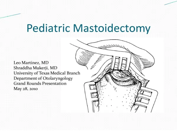
C.S.O.M.: Mastoidectomy. Dr. Vishal Sharma. Schwartze’s Cortical Mastoidectomy. Middle ear cleft (Right). Cortical Mastoidectomy. Boundaries of cavity. Superior: Dural or Tegmen plate Anterior: Posterior wall of external auditory canal Inferior: Digastric ridge

ashanti
Download Presentation

C.S.O.M.: Mastoidectomy

An Image/Link below is provided (as is) to download presentation Download Policy: Content on the Website is provided to you AS IS for your information and personal use and may not be sold / licensed / shared on other websites without getting consent from its author. Content is provided to you AS IS for your information and personal use only. Download presentation by click this link. While downloading, if for some reason you are not able to download a presentation, the publisher may have deleted the file from their server. During download, if you can't get a presentation, the file might be deleted by the publisher.

E N D

Presentation Transcript


  1. C.S.O.M.: Mastoidectomy Dr. Vishal Sharma

  2. Schwartze’s Cortical Mastoidectomy

  3. Middle ear cleft (Right)

  4. Cortical Mastoidectomy

  5. Boundaries of cavity • Superior:Dural or Tegmen plate • Anterior:Posterior wall of external auditory canal • Inferior:Digastric ridge • Posterior:Sigmoid sinus plate • Medially:Lateral semicircular canal

  6. Normal temporal bone (right)

  7. Cortical Mastoidectomy

  8. Indications

  9. Coalescent mastoiditis & Masked mastoiditis • CSOM T.T.D. active refractory to antibiotics • Secretory otitis media refractory to antibiotics • Approach to:  Endolymphatic sac surgery  Facial nerve decompression  Vestibulo-cochlear nerve section  Translabyrinthine approach for C.P. angle  Cochlear implant surgery  Combined approach tympanoplasty

  10. Antiseptic dressing

  11. Draping

  12. Infiltration with Lignocaine

  13. Marking of incision

  14. Wilde’s post-aural incision

  15. Incision deepened till periosteum

  16. Musculo-periosteal flap elevated

  17. Bezold’s abscess

  18. Aspiration of pus

  19. Drainage of abscess

  20. Drainage of abscess

  21. Corical mastoidectomy begun

  22. Exposure of mastoid antrum

  23. Widening of aditus

  24. Aditus widened

  25. Final Cavity (right)

  26. Final Cavity (left)

  27. Drain put in mastoid cavity

  28. Mastoid dressing

  29. Healed post-aural scar

  30. Treatment for Attico-antral disease

  31. Medical Treatment Topical ear drops + frequent suction clearance Indications: • Early disease with shallow retraction pocket • Only hearing ear with cholesteatoma • Elderly patients • Pts who are not fit for surgery under G.A. • Pts who can regularly come for follow up

  32. Surgical Treatment Canal Wall down: • Attico-antrostomy • Modified Radical Mastoidectomy (MRM) • Radical Mastoidectomy Canal Wall up: • Combined Approach Tympanoplasty (CAT)

  33. Mac Ewan’s triangle

  34. Canal Wall Up Mastoidectomy

  35. Canal Wall Down Mastoidectomy

  36. Middle ear cleft

  37. Attico-antrostomy

  38. Modified Radical Mastoidectomy

  39. Pre-operative Anatomy

  40. Cortical Mastoidectomy done

  41. Modified Radical Mastoidectomy

  42. Surgical Steps

  43. Perform cortical mastoidectomy • Lower facial ridge & break facial bridge • Remove cholesteatoma & granulations from mastoid air cells & middle ear cavity • Preserve healthy mucosa, T.M. remnant & ossicles • Mastoid cavity & E.A.C. become a single cavity seperated by middle ear cavity • Perform tympanoplasty. Do concho-meatoplasty.

  44. Antiseptic dressing

  45. Draping

  46. Infiltration

  47. Infiltration

  48. Marking of incision

  49. Wilde’s post-aural incision

  50. Wilde’s post-aural incision

More Related