Download
1 / 19
190 likes | 211 Views
This study explores the anatomy and physiology of the human eye and the effects of drugs on its functioning. It focuses on the regulation of pupil diameter and accommodation for near and far vision. The study also discusses the administration of drugs through topical, local, and systemic routes, as well as the use of ophthalmic anesthetics, dilating drops, and miotic agents. Additionally, the study examines the pathophysiology of glaucoma and its symptoms.

E N D
Study of the action of Drugs on Human EyesLab-7 WROOD SALIM, Msc. LABORATORY OF PHARMACOLOGY, UNIVERSITY OF AL-MUSTANSYRIA 2017-2018
Eye is specialized sensory organ that mediate vision. The eye focuses images from external environment onto retina & convert them into electrical signals which then recognized by the brain.
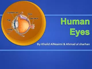
Anatomy & Physiology of the Eye Figure 1: The composition of the human eye
The main compartments of the human eye (as shown in figure 1) • 1- Iris: That involves: Circular muscle (Muscarinic receptors) and Radial muscle (Alpha-receptors). • 2- Lens: Attached to the ciliary body by ligaments. • 3- Ciliary body: that involves • Ciliary epithelium (B2 receptors): responsible for secretion of aqueous humor. • Ciliary muscle (M receptors): responsible for near or far vision • 4- The cornea is the transparentfront part of the eyethat cover the iris, pupil, and anterior chamber. • 5-The anterior chamber:is the fluid-filled space inside the eye between the iris and the cornea. Aqueous humor is the clear fluid that fills the anterior chamber.
Pupil Diameter The pupil is the hole in the center of iris. The diameter of the pupil (pupil size) & hence the amount of light entering the eye is regulated by two anatomically innervated set of smooth muscles: 1- radial muscles which innervated by adrenergic fibers (containing alpha1-receptors). 2- circular muscles which innervated by cholinergic fibers (containing M3-receptors). Notes:Miosis: is due to either contraction of circular muscle or relaxation of radial muscle. Mydriasis: is due to either contraction of radial muscle or relaxation of circular muscle
Human eye anatomy Mydriasis Miosis • Figure 2:Regulation of the amount of the light
Accummodation for Near & Far Vision Ciliary body involves ciliary muscle and ciliary epithelium Ciliary muscle (M receptors): responsible for near or far vision. M-agonist → Ciliary M. Contraction → Lens contraction → near vision Anti-Muscarinic → Ciliary M. Relaxation → Lens relaxation → far vision Contraction of ciliary muscle in response to cholinergic activation (M3 receptors) causes these suspensory ligaments to relax allowing the lens to become more convex & thusimproving the focus for closer objects. . relaxation of the ciliary muscle (e.g. by antimuscarinic agents) the suspensory ligaments will be stretch allowing the lens to become more flat so that improving the focus for farther objects. . Drugs that antagonized accummodation for near vision termed cycloplegics, they are exclusively muscarinic antagonists. sympatholytic agents do not alter accummodation for near vision since there are no adrenergic receptors in the ciliary muscle.
Human eyes accommodation Figure 3: The contraction and relaxation of the lens
Drugs and human eye • Topical administration • Eye drops • Principally absorbed through the cornea • Short drug-eye contact time • Eye ointments • Allow a prolonged contact time • Eye lotions • Used for irrigation • Local injections and systemic treatment • Physiological barriers limit systemically administered drug penetration to the eye • Ex. acetazolamide for severely raised intraocular pressure
Drugs and human eye • Ophthalmic anesthetics • Ophthalmic anesthetics are agents that act locally to block pain signals at the nerve endings in the eyes • Anaesthetic drops: • Initial assessment of minor trauma • Removal of conjunctival and corneal foreign bodies • In surgery • Example: • PropracaineHydrochloride 0.5% (Alcaine) • Tetracaine 0.5% • Side effects: • Allergy: local or systemic
Drugs and human eye • Dilating Drops (mydriatic medications) • Mydriatics are used to enlarge the pupil for eye examinations • Used in diagnosis and surgery • 1- Parasympathetic antagonists (parasympatholytics) • Paralyzing the iris sphincter muscle • Make the pupil larger and paralyze the muscle involved in focusing of the lens (accommodation) • Blurry eyes especially for up close (reading, near play) • Tropicamide: (Mydriacyl) 0.5% and 1%. Action up to 6 hours • Cyclopentolate: (Cyclogyl) 0.5%, 1% and 2%. Action up to 24 hours • Homatropine: 2% and 5%. Action: 2-3 days. • Atropine: (Atropisol) Drops 0.5% or 1%, ointment 1%. Action: 1-2 weeks • 2- Sympathetic agonists (sympathomimetics) • Stimulate the iris dilator muscle. • Phenylephrine: 2.5% and 10%. Action 3-6 hours.
Drugs and human eye • Miotic agents • Dapiprazole (α1-antagonist) • Pilocarpine (M3 agonist)
GLAUCOMA • Disease of the eye in which fluid pressure within the eye rises • May lead to vision lose • Affects both eyes • Symptoms :Loss of peripheral vision Sensitivity to light and glare • Problems with night vision ,and Blurred vision • Characterized chiefly by an increase in IOP above 21 mmHg & may be as high as 70 or 80 mmHg during the attack
Pathphysiology of Glaucoma • The aqueous humor is a transparent, gelatinous fluid. It is secreted from the ciliary epithelium. • In glaucoma, aqueous humor builds up and increases pressure within the eye • Ciliary Epithelium (B2-Receptors) Responsible for secretion of aqueous humor. • Ciliary muscle contraction(M3 agonist) → Increases flow → Decreases IOP. • Ciliary muscle Relaxation → Decreases flow → Increases IOP (Glaucoma).
glaucoma • Types of Glaucoma • Open-Angle Glaucoma • The most common form of glaucoma, 90% of all glaucoma cases • Caused by the slow clogging of the drainage canals, increased IOP • Develops slowly • “Open-angle” means that the angle where the iris meets the cornea is as wide and open as it should be • Angle-Closure Glaucoma • Caused by blocked drainage canals, sudden rise in IOP • Has a closed or narrow angle between the iris and cornea
glaucoma • Diagnosis • Tonometry: eyeball pressure is measured. High Intraocular pressure (IOP) may suggest glaucoma • Optic Nerve Examination: check for damage • Visual Field Examination: a patient's visual field (area in front) will be mapped to check for visual loss • IOP is the pressure caused by the fluid inside the eye that helps maintain the shape of the eye • IOP ranges from 10 - 21 mm Hg
glaucoma • Treatment • There is no cure for glaucoma and if the optic nerve is damaged, it cannot be fixed • The progression of glaucoma can be controlled, by lowering IOP • Increase the drainage • Decrease production • Drugs that reduce the production of aqueous humor: beta-blockers, alpha-adrenergic agents, carbonic anhydrase inhibitors • Drugs that increase the outflow of aqueous humor: prostaglandins, prostamides • Surgery to improve the outflow of aqueous humor • Implantation of a device to help drain fluid within eyes
OBJECTIVES At the end of the practical class the student shall be able to: 1. Instill drugs carefully into the volunteer eye by the pouch method without injuring the cornea. 2. Study the effects of drugs on the eyes
Methods • Place one drop of the agents in the following table into on eye and check for the parameters mentioned in the following table: