Download
1 / 15
150 likes | 335 Views
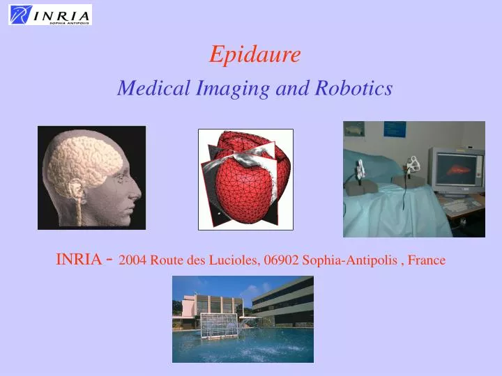
Epidaure Medical Imaging and Robotics. INRIA - 2004 Route des Lucioles, 06902 Sophia-Antipolis , France. Epidaure Team. Between 25-30 members including 5 permanent researchers : N. Ayache, H. Delingette, M.A. Gonzalez-Ballester, G. Malandain, X. Pennec

E N D
Epidaure Medical Imaging and Robotics INRIA - 2004 Route des Lucioles, 06902 Sophia-Antipolis , France
Epidaure Team • Between 25-30 members including • 5 permanent researchers : N. Ayache, H. Delingette, M.A. Gonzalez-Ballester, G. Malandain, X. Pennec • 2 engineers: E. Bardinet, M. Traina • 14 PhD Students (engineers and MDs) • Post-Docs • External Collaborators (Profs, MDs) • Trainees (Scientists, MDs) • Assistant : I. Strobant Epidaure
Objectives • Design et Develop new Software Tools to Improve the Impact of 3-D Bio-Medical Images. • Participate to theirValidation andTransfer through Medical and Industrial Partnership, and also through transversal actions. Epidaure
Current Situation ofMedical Imaging • Research: prevision of increase 30%/year until 2010 (x10) • Constant stream of better imaging&signal modalities • Emerging new modalities • Provides higher and higher resolution complementary information (spatial/temporal, anatomical/functional) • New image-guided therapies (includes MIT, robotics, cell and gene therapies) • Possibility to quantify the effect of a new medicine • Flow/quantity of information too high to allow optimal exploitation by simple visual examination Epidaure
MRI CT-Scanner Ultrasound Scintigraphy Some “Classical” Imaging Modalities Density of X-Ray absorption Density and structure of Protons Variations of Acoustic Impedance Density of injected isotopes Epidaure
Multidimensional Images Temporal Evolution of Multiple Sclerosis (collaboration with Harvard, CHU-Pasteur & QuantifiCare) 3 spatial dimensions (MRI-T2) Epidaure
Multispectral Images T1 T2 • Same patient, various MR sequences. PD Gd Flair Epidaure
Some New/Complementary Imaging Techniques • Optical imaging technologies • OCT (optical coherent tomography), Confocal imaging, auto/induced Fluorescence Imaging • Endoscopic imaging • Molecular imaging (molecular probes) • Tissue Biomechanics from Ultrasounds • NIR Topography/Tomography, • Laser Doppler, Thermoacoustics, Terahertz imaging, etc… • Histopathology images Epidaure
In Vivo Microscopic Confocal Imaging Source: Mauna Kea Technologies Reflectance imaging Depth of Observation up to 100 microns Fluorescence imaging Epithelium Lateral Resolution: 2 -- 4 mm Axial Resolution: 8 -- 20 mm - Confocal flexible microscope- High resolution optical sections of tissues 160 microns Epidaure
Why Medical Image Analysis? • Better individualised diagnosis • quantitative and objective measurements • from various sources of images/signals • acquired at various scales • Better individualised therapy • planning before • control during • evaluation after Epidaure
Epidaure Some Generic Research Topics • Restoration • Physics based Segmentation • Registration and Fusion • Statistical Shape Analysis • Atlas construction • Cardiac Motion Analysis • Functional MRI • Surgery Simulation • Biomechanical models • Physiological models • Visual/Haptic Interactions • Coupling with robotics... Epidaure
Some Current Clinical Projects • 5. Confocal Imagery • Inserm-U455 (Toulouse), TGS, • Mauna Kea Technologies, • 6. Functional MRI • CEA-SHFJ, Leuven, Odyssée • 7. Surgery Simulation • Ircad (Strasbourg), Mentice • Harvard Medical School • 8. Image-Guided Radiotherapy • IGR, Curie, CAL, DOSIsoft • 1. Multiple Sclerosis • Harvard Medical School, CHU-Pasteur, QuantifiCare • 2. Image-Guided Neurosurgery • Roboscope, • 3. Histological Atlases • Pitié-Salpêtrière, Medtronic • Qamric, Mapawamo • 4. Cardiac Motion • Johns Hopkins, Philips, GEMSE, ICEMA Epidaure
Conclusion • Analyse des images bio-médicales • recherche très active : recalage, mouvement, simulation, visualisation, indexation, segmentation, morphométrie, segmentation, statistiques, robotique, etc. • Nouvelles applications pharmaceutiques • mesures locales et dynamiques de l’efficacité de nouvelles molécules, de nouvelles thérapies (génique, cellulaire, etc.), • mesures quantitatives et objectives Epidaure
Medical Imaging and Robotics Design and Development of new Tools in Medical Image Analysis and Simulation to Improve Diagnosis and Therapy. EPIDAURE RESEARCH AXES COLLABORATIONS • General Electric MS, • Philips MS, • Mauna Kea Tech., • Medtronic, • Mentice, • Noesis, • Nycomed, • Philips MS, • QuantifiCare • Sanofi, • Siemens, • etc. • Extraction of quantitative parameters (shapes, textures) • Image Registration (temporal, multimodal, multipatients, etc.) • Construction of anatomical, histological and functional atlases from images • Morphometry (Statistics on shapes and intensity) • Analysis of cardiac motion • Virtual patients and surgery simulation (Visual and haptic feedback) • Image-Guided Surgery and Augmented Reality • Coupling medical imagery and medical robotics (with Chir) Epidaure
General References • Inria Reports and References on line • http://www-sop.inria.fr/ • Journals • Medical Image Analysis (Elsevier) • Computer Aided Surgery (Wiley) • Transactions on Medical Imaging (IEEE)... Epidaure