Download
1 / 32
400 likes | 842 Views
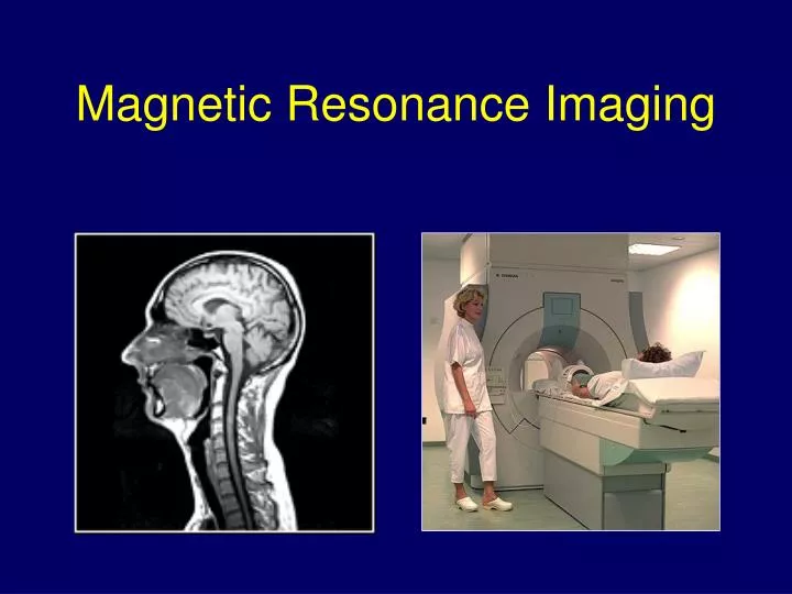
Magnetic Resonance Imaging. Magnetic field gradients. Gradient echo. T 2 * envelope. M xy. Time. Equal areas. Gradient Amplitude. Gradient echo. Slice selection using gradients. Sample. B z. B (x 1 ). B (x 2 ). x. x 1. x 2. Slice selection.

E N D
T2* envelope Mxy Time Equal areas Gradient Amplitude Gradient echo
Slice selection using gradients Sample Bz B(x1) B(x2) x x1 x2
Gradients can be combined to produce gradients in arbitrary directions
Imaging in gradients Sample Bz B(x1) B(x2) x x1 x2 Signal (spectrum) Frequency w1 w2 PROJECTION
Basic reconstruction X-ray beam is attenuated forming a projection
The projection tells you that the beam was attenuated with equal probability anywhere along its path
Projection Reconstruction • Diffusion imaging • Radial Spin Echo and TSE Scan PR • Low sensitivity to motion • Low sensitivity to susceptibility artifacts
Projection Reconstruction • Sodium Imaging • Human Skeletal Muscle: Sodium MR Imaging and Quantification-Potential Applications in Exercise and Disease • Constantinides, Gillen, Boada, Pomper, Bottomley, • Radiology
K-space Image
K space Units: rad/ m Increasing spatial frequency
Spin-warp pulse sequence Spin-warp k-space trajectory ky RF C 1 B 2 3 Gslice 1 2 3 kx Gphase A Time n Gread n Signal A B C With MRI we can control how we sample k-space
Spin warp imaging Wicklow, Washington University, St. Louis
Roger Ordidge, UCL
Motion artefacts Anesthetic Steve Keevil UMDS
Fast spin echo T2 weighted
Turbo/Fast field echo T1 weighted
TFE for Phase v T2* contrast Modulus Phase
T1Contrast: MPRAGE & IR-TSE IR-TSE: 0.25 x 0.25 x 1.5 mm3 MPRAGE: 0.5 mm isotropic (11 mins) 384 x 384 x 200
Gradient safety: Peripheral nerve stimulation Numerical Simulations • PNS is associated with hotspots of electric field (E) or current density (J) • Get “hot-spots” of |E|/|J| in low/high conductivity tissues. Head-centred coronal slice y = 0 E J J
Gradient field: Acoustic noise • One of the biggest challenges at high field • Acoustic noise • Ear defenders and foam to reduce bone conduction • Gradient system decoupled from bed (and room) • Modification of gradient waveforms • Novel gradient coil designs Conventional Waveform Minimum slew rate waveforms