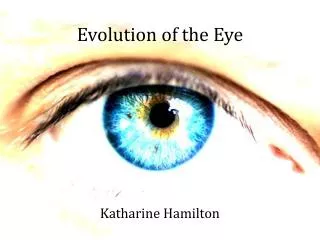
DEVELOPMENT OF EYE
DEVELOPMENT OF EYE. DEVELOPMENT OF EYE. Eyes are derived from four sources Neuroectoderm of fore brain retina, posterior layers of iris, optic nerve Surface ectoderm of head Lens& corneal epithelium Mesoderm between above layers Fibrous & vascular coats Neural crest cells


DEVELOPMENT OF EYE
E N D
Presentation Transcript
DEVELOPMENT OF EYE Eyes are derived from four sources • Neuroectoderm of fore brain retina, posterior layers of iris, optic nerve • Surface ectoderm of head Lens& corneal epithelium • Mesoderm between above layers Fibrous & vascular coats • Neural crest cells choroid, sclera & corneal endothelium
OPTIC PRIMORDIA • The developing eye appears in the 22-day embryo as a pair of shallow grooves on each side of the forebrain.
DEVELOPMENT OF OPTIC CUP & LENS VESICLE • With closure of neural tube these grooves will form outpocketings of the forebrain, optic vesicles • On contact with surface ectoderm, these vesicles induce changes for lens formation, shortly there after the vesicles invaginate and form the double walled optic cup
Cont… • The inner and outer layers of optic cup are separated by intraretinal space, soon the lumen disappears and the layers fuse • Rim of the cup in folds around the lens vesicle
Cont… • Invagination is not restricted to the central part of optic cup but also involves part of inferior surface that forms the retinal / choroid fissure
Cont… • Vascular mesenchyme give rise to hyaloid vessels which supplies the inner layer of optic cup, lens vesicle & mesenchyme in optic cup
Cont… • As the edges of retinal fissure fuse these vessels are enclosed with in primordial optic nerve • Distal parts of these vessels degenerate while proximal parts persist as central artery & vein of retina
Cont… On contact of optic vesicle with surface ectoderm, the cells of surface ectoderm elongates and form the: • Lens placodes • Lens pit • Lens vesicle
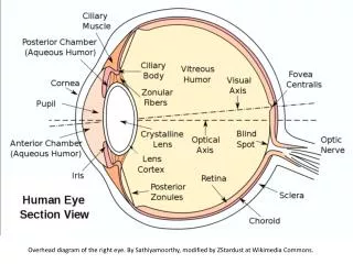
DEVELOPMENT OF RETINA Retina develops from the walls of optic cup • Outer layer Pigment epithelium of retina • Inner/Neural layer Posterior 4/5th pars optica retinae (NeuroEp) Intraretinal space; disappears when two layers fuse Rods & Cones Mantle zone
On the surface are the axons of ganglion cells that grow in optic stalk to form optic nerve fiber layer • Neural retina is inverted • Light impulses pass through most layers of retina before they reach photoreceptors
pars ceca retinae The anterior 1/5th of the inner layer of retina (pars ceca retinae ) divides into: • Pars iridica retinae ---- inner layer of iris • Pars ciliaris retinae ---- ciliary body The region b/w the optic cup and the overlying surface epithelium is filled with loose mesenchyme.
CILIARY BODY • Is the part of anterior 1/5th of the inner layer • Medial surface projects towards lens forming folds --- ciliary processes • The pigmented outer portion of ciliary epithelium is derived from outer layer of optic cup & is continuous with retinal pigment Ep. • The nonpigmented portion of ciliary Ep. represent the anterior prolongation of neural retina and consist of single layer of epithelium • The ciliary muscle develops from the mesenchyme of optic cup---- smooth muscle for focusing the lens
DEVELOPMENT OF IRIS Develops from rim of optic cup that grows inwards & partly covers the lens Iris is formed by: • The pigment containing external layer continuous with the pigment epithelium of ciliary body & neural retina • The unpigmented internal layer of the optic cup C.T frame work is derived from neural crest cells that migrate into iris The dilator pupillae & sphincter pupillae muscles develop in the surrounding mesenchyme and are derived from the underlying ectoderm of the optic cup
DEVELOPMENT OF LENS LENS VESICLE: Derivative of surface ectoderm Anterior wall : Cuboidal Ep (sub capsular lens Ep.) Posterior wall : Tall Columnar Ep--- nuclei dissolute cells elongate anteriorly & form primary lens fibers which gradually obliterate the lumen of lens vesicle Equatorial zone : The rim of lens, midway between anterior & posterior poles of lens Cells are cuboidal first, elongate, lose nuclei & form secondary lens fibers which re continued to form during adult hood Nutrition of lens: Initially by distal part of hyaloid artery which degenerates; later it depends upon aqueous humor & Vitreous humor
Tunica vasculosa lentis: capsule of lens Anterior part forms pupillary membrane Both degenerate when hyaloid A. degenerates However the lens capsule produced by anterior lens Ep. & lens fibers persist
DEVELOPMENT OF VITEROUS BODY • Forms within the cavity of optic cup • Mesenchyme not only surrounds the eye primodium but also invades the inside of optic cup by way of choroidal fissure • Here it forms hyaloid vessels which supplies posterior surface of lens& forms a vascular layer on inner surface of retina • Forms delicate network of fibers between retina & lens • Interstitial space fills with delicate gelatinous substance forming vitreous body • Hyaloid vessels disappear leaving hyaloid canal only
DEVELOPMENT OF AQUEOUS CHAMBERS • ANTERIOR CHAMBER develops from clefts that appear in the mesenchyme between developing lens & cornea • POSTERIOR CHAMBER develops from the space that forms in mesenchyme posterior to developing iris & anterior to developing lens • When pupillary membrane disappears & pupil forms the anterior & posterior chambers of the eye are able to communicate with each other. The clear aqueous humor circulates from posterior to anterior chamber and provides nutrients for cornea and lens. From anterior chamber the fluid passes through the scleral venous sinus (canal of Schlemm) at the iridocorneal angle where it is resorbed into blood stream. This drains the anterior chamber into venous system
DEVELOPMENT OF CHOIRD, SCLERA & CORNEA • Under the inductive influence of retinal pigment epithelium the mesenchyme surrounding the optic cup differentiates into an inner vascular pigmented layer, the choroid & outer fibrous layer, the sclera • Choroid becomes modified to form cores of ciliary processes containing delicate capillaries & fine C.T • Sclera is continuous with corneal stroma • Lens vesicle induces the formation of cornea by transforming surface ectoderm into transparent, multi layered,a vascular cornea
CORNEA is formed from three sources • External corneal Ep.--- surface ectoderm • Mesenchyme (stroma) --- continuous with sclera • Internal corneal endothelium--- neural crest cells