Download
1 / 19
240 likes | 513 Views
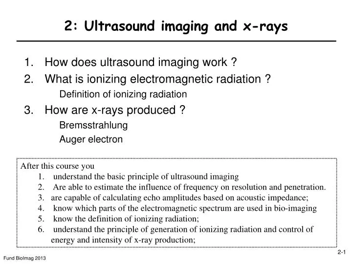
2: Ultrasound imaging and x-rays. How does ultrasound imaging work ? What is ionizing electromagnetic radiation ? Definition of ionizing radiation How are x-rays produced ? Bremsstrahlung Auger electron. After this course you understand the basic principle of ultrasound imaging

E N D
2: Ultrasound imaging and x-rays • How does ultrasound imaging work ? • What is ionizing electromagnetic radiation ? Definition of ionizing radiation • How are x-rays produced ? Bremsstrahlung Auger electron • After this course you • understand the basic principle of ultrasound imaging • Are able to estimate the influence of frequency on resolution and penetration. • are capable of calculating echo amplitudes based on acoustic impedance; • know which parts of the electromagnetic spectrum are used in bio-imaging • know the definition of ionizing radiation; • understand the principle of generation of ionizing radiation and control of energy and intensity of x-ray production;
What do these have in common ? Ultrasound transducer US scanner Orca Ship HumanHair Single Crystal Bat Microscopic view of scanhead
2-1. What are the main fates of US waves in matter ? 2. Refraction 3. Scatter 1. Attenuation Sound wave bends as it hits an interface at an oblique angle Sound wave dispersed in all directions Sound wave travels through the substance but loses energy I(x) Reflection (echo formation) is key to imaging 4. Reflection Attenuation coefficient a [dB/(cm Mhz)] a is usually given in dB: dB=10logI(x)/I0 [3dB=2fold increase in I(x): 100.3=2] Typically a~0.5dB/(cm MHz) → 6MHz signal will lose 3dB per cm of travel (2 fold loss in wave energy) Sound wave bounces back to probe
What is the basic principle of US imaging ? • The basic principle of imaging using sound waves : • Emit sound pulse (length [1-5 µs] is a multiple of cycle time 1/f) • Measure time and intensity of echo • Reconstruct using known wave propagation velocity c • UItrasound: frequency f=1-20MHz (not 20kHz) • Sound wave propagation velocity c [c=lf] ~330m/s (air) = 0.33 mm/µs ~1.45-1.6 mm/µs (tissue) (1cm~7µs)(increases with density r, bone ~ 4 mm/µs) Distance of tissue boundary from probe (transducer) transducer Distance=speed x time/2
What determines the resolution in US imaging ? x Overlap: No Gap, No separate echoes Gap: Separate echoes T1=2x1/c DT=T1-T2=2Dx/c T2=2x2/c min. echo separation, e.g., DT ≥ 2 Dt Pulse duration Dt = N/f • Wavelength l determines minimal resolution • To have defined frequency:Pulse length = N/f l • Separation of return echoes, e.g. DT > 2 pulse length • 1. Resolution • increases with f • 2. Penetration (cf. attenuation) • decreases with f Free lunch
When does an acoustic echo occur ? Acoustic impedance and reflection ratio Acoustic impedance Z • Definition: • Z= rc [kg/m2s=rayls] Amount of reflected wave energy Iref=I0RI At interface between objects with different acoustical properties Probability of reflection + transmission is = 1: Reflection coefficient Transmission Z1 Z2
What are the reflection coefficients RI between tissues ? US shadow due to gallstone Reflection by solid material e.g. bone-tissue interface Shadow formation: ~45% of energy transmitted 100% 45% (TI=1-RI) 45% 20% Dolphin fetus bone
What is the optimal choice of US frequency ? The optimal frequency decreases with tissue depth and with increasing absorption Resolution SNR • SNR: • Signal returned from an echo-generating tissue interface at distance x from transducer Resolution: Dxdecreases with increasing frequency f : 1/f Resolution f =0 f0=1/(2ax) is constant How critical is the choice of f0 ? f: US frequency (experimental parameter) a: attenuation coefficient (tissue parameter) Find the optimal f … Maximize f·S D(fS)<20%: 4-fold range of f Maximum is where derivative with respect to f is zero is constant f0
Examples High-resolution US at the surface: Skin, subcutaneous tissue Low-resolution US of deep tissue Epidermis Loose connective tissue and subcutaneous fat Muscle interface Muscle fibres interface Bone Liver metastases
Ex. 3-D US Imaging & Contrast agents Contrast agents: gas-filled Bubbles • 3D US Physical Principle: • the transducer is moved during exposure (linear shift, swinging, rotation) • received echoes are stored in the memory • the image in the chosen plane is reconstructed mathematically Gas : most contrast(plus resonance and higher harmonic imaging) (see tiny Z → total reflection, RI~1) Umbilical cord Kidney perfusion (mouse)
How can Ultrasound detect moving blood ?Doppler effect stationary Source moving with v0 • Motion (Doppler): Frequency shift fD of moving tissue, results in shifted US frequency (demodulation for detection) • (where is this also used?) In a period T, source moves closer by v0T lf=(c-v0)T l=cT Doppler frequency shift fD c: speed of US, e.g. 1500 m/s lr=(c+v0)T v0: speed of source, e.g. 50 cm/s f0: frequency of moving source, e.g. 5MHz a: Rel. angle at which blood is moving Example: fD= 2·5·106 [Hz] 0.5 [m/s]/1500 [m/s] ~ 3kHz ~ 0.05% of f0 Doppler US of internal carotid artery stent
X-raysThe beginnings of biomedical imaging Wilhelm Röntgen, Nobel Prize Physics 1901
2-2. Basis of x-ray imaginguseful relationships Electromagnetic radiation c = ln(c = speed of light = 3∙108m/s) • E = hn=hc/l(h = Planck’s Constant) h= 2pe-34Js = 4e-18keVs 1eV = energy of e- in acquired in 1V electric field • E = hc/l = 1.2keV/nm NMR 10 -800 MHz
With which elements of matter does EM radiation interact mainly ? (in imaging mainly with electrons) Electron binding energy • Binding energy • decreases with shell distance • increases with Z • (Why?) • Lowest K-shell binding e- energy: • EKmin = 13.6eV (1H) • hn > EKmin: ionizing • hn < EKmin: non-ionizing • Electron (some useful constants) • me = mass = 9e-31kg • qe = charge = 1.6e-19 C (As) • Rest energy mec2 = 511 keV Ionizing radiation is above 13.6 eV
What is ionizing radiation ? 13.6eV Non-Ionizing Ionizing
2-3. How are x-rays generated (scheme) ? • Negatively charged cathode = electron source • Electrical current (filament current) heats up the cathode (why is that necessary ?) • Electrons are liberated and accelerated by electric field (Energy of e-= qDV) • Anode = metal target (tungsten) • accelerated electrons hit anode generate X-rays • (tube current with voltage difference up to 150 kV) • Intensity of beam = Power/Area • Number of X-rays (proportional to tube current) • Energy of X-rays hn(proportional to voltage)
- Emission of x-rays I: What is Bremsstrahlung ? pi Consider the interaction of e- with stationary atom as collision : pi=pf+pphoton • Elastic scattering: Probability ~ Z2/Ee-2 • Inelastic scattering: n release Probability ~ Z2 No info on directionality of radiation (but maximum energy is defined, how?) • Coulomb: • a ~ qeZ/mer2 • PBrems = qe2a2/6pe0c3 pphoton pf Max. Energy: Eei High Z: Tungsten is a good target Decreasing energy
Emission of x-rays II: What are Characteristic (fluorescent) X-rays ? Auger emission The excited atom can also reduce energy by liberating an additional e- (Auger e-): • Impacting e- liberates inner shell e- • Atom is excited (higher energy state) • Vacancy • Filled by outer shell electron (cascading) • Emission of characteristic x-ray