Dermatomyositis
Dermatomyositis. Floranne Ernste , MD The Myositis Association Annual Conference Bloomington, MN Septem ber 2019. Disclosures. Octapharma : clinical trial support Genentech: clinical trial support Off-label use: Almost all medications discussed here for treatment are not FDA-approved.

Dermatomyositis
E N D
Presentation Transcript
Dermatomyositis FloranneErnste, MD The Myositis Association Annual Conference Bloomington, MN September 2019
Disclosures • Octapharma: clinical trial support • Genentech: clinical trial support • Off-label use: • Almost all medications discussed here for treatment are not FDA-approved.
Learning Objectives • Gain an understanding of the definition of dermatomyositis and how we diagnose it • Appreciate how different groups of people are affected • Recognize varied treatment options • Discuss short and long term prognosis
Dermatomyositis: what is it? • An autoimmune disease that causes inflammation in your skin and muscle tissue • Take a step back: what is autoimmune disease? • When the immune system goes awry. • How? Your immune system loses “tolerance” to your body’s tissues and certain cells such as your “B cells” and “T cells” and other cells start fighting against your tissues by mounting an inflammatory response.
How it may all start... Trigger(s): UV light exposure, certain infections, cancer Abnormal Immune System Response (your genes inherited determines your immune response) Organ/Tissue Damage: i.e. skin, muscle, lung, heart, joints Clinically manifests as rashes, muscle weakness, joint pain/swelling
Dermatomyositis: what is it? • 3 main types of DM: • Amyopathicdermatomyositis • Rashes without muscle involvement for 6 mos. or longer • Hypomyopathicdermatmyositis • Rashes with minor abnormalities in muscle testing, but not clinically weak • Dermatomyositis • Rashes and muscle weakness • Juvenile Dermatomyositis • Dermatomyositis in patients who are diagnosed at 16 years or younger
How Common is IT? • Not very common • Olmsted County Data, population based study • Incidence of 9.63 per million; Prevalence of 21.42 per 100,000 • Peak age is 45 to 65 years for adults; 5 to 15 years for juvenile DM • Female-to-male ratio is 2-3:1 • In US, African Americans may be more commonly affected than Caucasians in a ratio of 3-4:1 Bendewald MJ, Wetter DA, et al. Arch Dermatol. 2010
Skin manifestations of dermatomyositis • Heliotrope rash: this is a lilac or purple rash affecting the eyelids mainly, but also above and below the eyes • Gottron’s papules: purple flat or raised bumps over knuckles, fingers, elbows, knees
Skin manifestations of dermatomyositis • V-sign: violaceous rash over neck and chest • Shawl-sign: violaceous rash over shoulders, upper arms • Nailfold abnormalities: cuticule overgrowth, dilated capillary loops
Skin manifestations of dermatomyositis • Calcinosis: hard skin lumps under skin, subcutaneous tissues, intramuscular tissue: more common in juvenile DM and a small percentage of adult DM
Muscle manifestations of dermatomyositis • Weakness of proximal muscles (neck/upper arms/thighs/hips) occurs gradually over several months. • Can start with difficulty getting in/out of vehicles, climbing stairs, raising your arms overhead with a heavy object in hand, lifting a gallon of milk, carrying your groceries, getting up off the toilet, can’t get up quickly after sitting on floor without help • Might feel a soreness in your muscles, rarely severe pain • Might notice less muscle tone/bulk, sometimes progresses to “atrophy” • You might start to notice difficulty swallowing bulky/dry foods, also known as “dysphagia”
What is happening at muscle tissue level? T2 weighted MRI of thighs
How do physicians diagnose dermatomyositis? • Physical exam: are there DM rashes present, muscle weakness? • Lab tests: Do you have elevated muscle enzymes such as creatinekinase, aldolase, AST (SGOT), ALT (SGPT), LDH • Electromyogram (EMG) • MRI of muscles (sometimes used) • Muscle biopsy
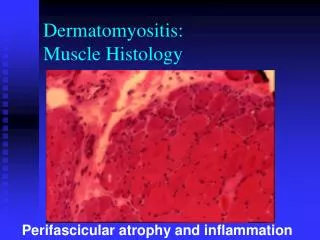
What do these test results look like? • Muscle biopsy • Perivascular infiltrate (inflammation) around muscle fascicle • Perifascicular atrophy* • Muscle microvasculature often involved • Necrotic and regenerating fibers seen http://www.neuro.wustl.edu/neuromuscular/pathol/inflammation.html
Learning Objectives • Gain an understanding of the definition of dermatomyositis and how we diagnose it • Appreciate how different groups of people are affected • Recognize varied treatment options • Discuss short and long-term prognosis
Muscle antibodies in dermatomyositis • In myositis patients, approximately 60-70% of adults and children have a myositis autoantibody • There are myositis specific antibodies and myositis associated antibodies • MSA are usually specific for PM/DM • MSA are usually mutually exclusive (not always in my experience). • Definition of MAA is broad: found in PM/DM, yet also found in other autoimmune rheumatic diseases. • May be part of an overlap syndrome such as SLE and scleroderma. • Can co-exist with other MSAs
How does DM affect different people? Patient 1 • 32 year old Hispanic male with itchy red rashes over face, neck,upper chest, arms, hands, fingernails, hips and thighs for 2 years. • Presented with significant hand arthritis with swelling of knuckles and wrists • Minimal muscle involvement, no significant weakness. Normal CK and other muscle enzymes. • Work-up revealed a positive MDA-5 (a myositis antibody)
MDA-5 positive antibody • Inflammatory arthritis may be severe, often appears “rheumatoid-like.” • Mild myositis. • Unique cutaneous characteristics • Mucocutaneous ulcerations • Tender palmar/digital papules, i.e. “inverse Gottron’s papules” Fiorentino D, Chung L, Zwerner J. J Am AcadDermatol. 2011;62:25-34.
MDA-5 antibody • First described 2009 in Japanese/South Korean cohorts of CADM, “clinically amyopathicdermatomyositis” • Targets melanoma differentiation association gene 5 protein, a cytoplasmic, viral RNA-specific helicase involved in innate immune response • Functions as an “anti-viral” sensor • Induces type I interferon responses • Can infection trigger this? • Associated with rapidly progressing ILD: 50% in Japanese cohort and 22% in USA Sato S, Hoshino K, Satoh T, et al. Arthritis Rheum 2009;60:2193 Sato S, Hirakata M, Kuwana M, et al. Arthritis Rheum 2005;52:1571-76. Fiorentino D, Chung L, Zwerner J, et al. J Am AcadDermatol. 2011;65:25-34.
How does DM affect different people? Patient 2 • 65 yo white male with fevers and weight loss for 6 months develops weakness and difficulty with swallowing. • His work-up was notable for high muscle enzymes, abnormal EMG. A muscle biopsy revealed dermatomyositis. • Additional work-up included a PET scan and then an upper endoscopy (EGD).
Cancer and Dermatomyositis • Cancer and myositis first reported in 1916 with DM and stomach cancer. • 25% of DM patients over age 50 may have cancer. • Cancer risk is greater in first 3 years of diagnosis (really highest in 1st year of dx). • Patients will usually have features of unusual weight loss, maybe fevers, rapid decline in physical function • Some cancers: Lung, breast, ovarian, colon, bladder Stertz G. Polymyositis. Berl Klin Wochenschr. 1916;53:489. Gerami P, Schope J, McDonald L, et al. J Am AcadDermatol. 2006;54(4):597-613. Callen JP. Dermatomyositis and malignancy. Clin Rheum Dis. 1982;8:369-81. Oldroyd et al. Clinical Medicine 2017.
How are different people affected? Patient 3. • 57 yo white woman presented to clinic with rashes over face, chest, hands inflammatory arthritis involving small joints of hands and dyspnea • Skin biopsy revealed “vacuolar interface dermatitis” • Chest x-ray showed bi-basilar infiltrates • Within 2 weeks of presentation, admitted to ICU with acute respiratory failure • Intubated, started on treatment for myositis and lung disease, and after 2 weeks hospitalization, died
Dermatomyositis and pulmonary manifestations • At least 30% myositis patients have interstitial lung disease (ILD) • This may manifest as fatigue and dry cough, shortness of breath that gets worse with minimal activity • Patients also may have respiratory muscle weakness • Anti-Jo-1 antibody found in 50-75% myositis pts with ILD • Strong association of ILD with all anti-synthetase antibodies in myositis. • No correlation between extent and severity of muscle or skin disease and development of ILD. • Secondary pulmonary hypertension can occur due to chronic pulmonary vasoconstriction from hypoxemia.
Dermatomyositis and pulmonary manifestations • Onset of ILD variable: most of the time occurs at the same time of myositis diagnosis. • Course ranges from acute and fulminant ILD, chronic progressive, or asymptomatic (subclinical). • Pulmonary function testing reveals restrictive physiology (i.e. FVC ≤ 80%). • ILD subtype classified as non-specific interstitial pneumonia (NSIP)—most common, cryptogenic organizing pneumonia (COP), and usual interstitial pneumonia (UIP). • Chest imaging shows basilar abnormalities: reticular and ground-glass opacities with loss of lung volume, traction bronchiectasis • ILD leads to poor functional status in 30% of patients.
How are different people affected? Patient 4 • 7th grade girl with JDM since age 7 presents with painful subqcalcinosis of posterior thighs, left forearm, antecubitalfossa (elbow region), buttocks. • Hurts to sit down in class. • Past treatments: prednisone, methylprednisolone, IVIG, methotrexate,CellCept, rituximab, cyclosporine, and hydroxychloroquine. • Exam notable for indurated skin of bilateral thighs, hardened, painful. • Underwent several debulking surgeries to remove calcium deposits.
How are different people affected? Patient 4 T2 Weighted Coronal Image CT Cross section views
How are different people affected? Patient 4 • Treated with ultrasound and CT-guided aspirations and resection of posterior calcific masses. • Developed complications (5-10 years’ worth): • Chronic, infected fluid collections with sinus drainage • Numerous hospitalizations with multiple aspirations, resections and antibiotics’ courses • Eventually went off all medications for several years, then recurrence in 20s of severe calcinosis in a leg, now back on therapy
Calcinosis and dermatomyositis • Calciniosis occurs from deposition of hydroxyapatite calcium phosphate in the soft tissues. • Calcinosis is more frequent in pediatric age groups, such as JDM, occurring 30-70% of time (associated with aggressive disease and delay in dx), and in adults about 15-20%. • Calcinosis believed to result from intracellular accumulation of calcium secondary to a disruption in cell membranes from trauma/chronic inflammation. • Multiple patterns include superficial papules or nodules, deeper nodules or tumors in the dermis or subcutaneous tissue, or diffuse deposits along the myofascial planes which may form an extensive exoskeleton.
Dermatomyositis: how does it affect non-white groups? • Clinical characteristics: rashes may be subtle and not classic in appearance. • Higher risk of calcinosis in non-white children with JDM • CK is often higher in African and African-American populations than in white groups • Organ involvement may be more severe in African, African-American and Hispanic populations. • For example, interstitial lung disease often is more severe and refractory to standard treatment than in white patients • Highest rates of hospitalization in non-white DM patients • Mortality rates may be higher in women of non-white groups Pinal-Fernandez, Iago et al. 2017. Rheumatology, 56(6); 999-1007. Hochberg, MC, et al. 1983. Arthritis & Rheumatism, 26 (12); 1465-1471. Kwa, M., et al. 2017. Arthritis Care & Research, 69(9);1391-1399. Hoeltzel, MF, et al. 2012. Pediatric Rheumatology, 10 (Suppl 1).
Learning Objectives • Gain an understanding of the definition of dermatomyositis and how we diagnose it • Appreciate how different groups of people are affected • Recognize varied treatment options • Discuss short and long-term prognosis
Treatment of DM • Should involve a multi-specialty approach with therapy targeted to skin and/or muscle, physical therapy, occupational therapy. • There is a lack of large randomized-controlled trials evaluating efficacy of treatments used by experts. • Management of extra-muscular organ involvement such as lungs, cardiac is important. • Involvement of specialists in multiple medical or surgical disciplines: neurologists, rheumatologists, dermatologists, physical medicine, pulmonologists, cardiologists, general surgeons, orthopedic surgeons is important.
My approach to treatment • First line: Glucocorticoids (topical +/- oral)+ a steroid sparing agent: • Azathioprine (2 mg/kg) • Methotrexate (if no severe ILD) • Mycophenolatemofetil (standard dosing or 1500 mg twice daily). • Glucocorticoid dosing: pulse intravenous daily (3 days) for severe disease (i.e. severe weakness, dysphagia, progressive ILD). • Oral glucocorticoids with taper: 1 mg/kg for 4 weeks, taper by 1-2.5-5 mg every 2-4 weeks pending treatment response and tolerability. • Oral taper may slow or stop ~5-10 mg daily.
My approach to treatment • Anti-malarials may help skin disease such as hydroxychloroquine, sometimes not adequate alone, and not effective for muscle disease • Intravenous immunoglobulin (IVIG) may be used initially or as “bridge therapy” until maintenance immunosuppressives kick in. • IVIG 1-2 gram/kg of ideal body weight. • Some patients do not tolerate due to headaches, aseptic meningitis • Lower doses may be used.
My approach to treatment for severe ILD or other severe organ disease • IV or oral cyclophosphamide. • Data exists for improvement in Jo-1+ pts with ILD in small series of patients. • Cyclosporine A or Tacrolimus • Data exists in several small series of patients. • I have more experience with tacrolimus, twice daily dosing targeting a trough level of 5-20 ng/mL. • Monitor for hypertension, renal insufficiency, electrolyte derangements, tremor, peeling rashes. Marie I, Josse S, Hatron PY, et al. Arthritis Care Res (Hoboken); 2013; 65:800-8. Oddis CV, Sciurba FC, Strazl TE. Lancet. 1999;353:1762-63. Wilkes MR, Sereika SM, Fertig N, et al. Arth Rheum. 2005;52:2439-2446
My approach to treatment for refractory myositis disease • Rituximab 1000 mg X 2 and repeat every 6 months; doses may be reduced in subsequent infusions • “RIM” trial of refractory juvenile/adult IIM, didn’t meet endpoints, but 83% met definition of improvement. • Refractory IIM patients with strongly positive autoantibodies (i.e. Jo-1) may be more responsive to rituximab (shorter time to improvement). • Interestingly, autoantibody titers decrease after rituximab suggesting a correlation with clinical response. Oddis CV, Reed AM, Aggarwal R, et al. Arthritis Rheum.2013;65:312-24. Aggarwal R, Bandos A, Reed AM, et al. Arthritis Rheumatol. 2014;66:740-9
What about calcinosis? • Numerous therapies tried with varying or no success: • Calcium channel blockers (diltiazem, amlodipine) • Bisphosphonates (alendronate) • Colchicine • IVIG • Methotrexate • Steroids • Biologic medications • *Topical sodium thiosulfate (useful for small calcific deposits under skin) • IV sodium thiosulfate (experimental) • Surgical resection/debulking
Newer treatments on horizon • IL-6 blockers • JAK inhibitors • Tofacitinib-recalcitrant DM • Abatacept • Lenabasum for skin disease • Sifalimumab • Anti-IFN alpha monoclonal ab Tjarnlund A, Tang Q, Wick C et al. Ann Rheum Dis 2018;77:55-62
Physical therapy • Beneficial. • Effective on muscle performance, aerobic capacity and overall health. • Exercise may downregulate genes related to inflammation. • “If you don’t use it, you lose it.” • I try to engage patients to meet with physical therapist early for stretching exercises and submaximal strengthening exercises. • Good to prevent muscle damage leading to muscle atrophy, preserve joint range of motion. • Safety assessment may be required if falls are common and gait aids may be appropriate. MuntersAlemo L, Alexanderson et al. CurrRheumatol Rep 2014;16 (7) 429
How is treatment response assessed? Core set measures developed. • Manual muscle strength testing (MMT) • Functional assessment – HAQ,CHAQ, FI • Global assessment • Physician • Patient/parent • Assessment of extra-muscular activity - MDAAT/MITAX or CMAS • Muscle enzymes - CK, aldolase, AST, ALT, LDH Miller FW, Rider LG, Chung YL, et al. Rheumatology 2001;40:1262-73
My assessment of treatment response • Improvement or stabilization of muscle strength on physical exam. • Normalization of muscle enzymes: CPK, aldolase, LDH, AST, ALT. • I may check serial EMGs or muscle MRIs. • Improvement of other organ systems such as pulmonary and cardiac: • Serial PFTs • Serial chest imaging, preferably high resolution CT imaging. • Echocardiogram to screen for heart failure and pulmonary hypertension
Management of side effects and other concerns. • If active skin disease, the sun is no longer your friend. • Concern for UV radiation exposure may trigger or exacerbate skin disease; increase risk for skin cancer • Certain medications may enhance sunlight effect • Wear protective clothing (wide brimmed hat, long sleeves if appropriate) • Some patients buy UV photo-protective clothes, expensive • Wear sunscreen and reapply at least every 2 hours even on a cloudy day (SPF of 50 or higher)
Management of side effects and other concerns. • Screen for latent TB, HIV, chronic hepatitis B and C infections. • Check vaccination status including influenza, Shingrix and pneumococcal vaccines (PCV-13, PPSV-23). • Screen for diabetes, hyperlipidemia, hypertension, osteoporosis at baseline. • Use calcium/vitamin D when on steroids. • If osteoporosis develops, use anti-resporptive agent or other agent if appropriate.