Paraneoplastic Opsoclonus
931-1. Paraneoplastic Opsoclonus. History. In July 1986 a 58-year old woman presented acutely with nausea, vertigo, difficulty focusing and oscillopsia. A diagnosis of labyrinthitis was made

Paraneoplastic Opsoclonus
E N D
Presentation Transcript
931-1 Paraneoplastic Opsoclonus
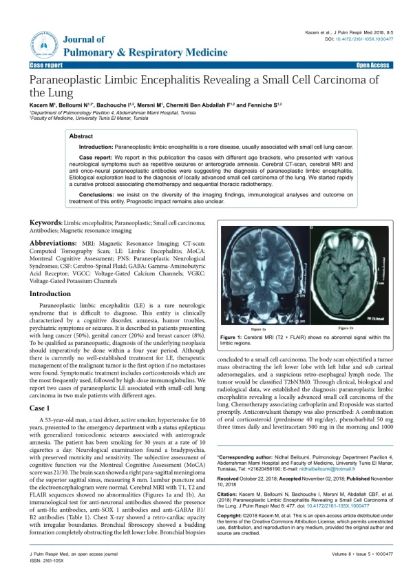
History In July 1986 a 58-year old woman presented acutely with nausea, vertigo, difficulty focusing and oscillopsia. A diagnosis of labyrinthitis was made Two later she was referred with persistent oscillopsia and the sensation that she was hanging upside down
Admission O.E extremely ill, vomiting mentally alert, intact speech + titubation, myoclonus, trunkal ataxia Blood test showed hyponatremia consistent with SIADH CFS: protein 54 mg/dL, 24 WBC 92% lymphs, multiple oligoclonal bands
Figure 8. Crebellum/Neurones. Anti-Ri antibody against neuronal nuceli.
Figure 11. Functional MRI during opsoclonus: activation is found in the cerebellum but not in the pontine brainstem in both patients. For better illustration, only the cerebellum and brainstem are shown. Increased activation is shown on individual brian images for the sagittal (s), coronal (C), and transversal (T) slices at the level of highest activation in the midline cerebellum for Patient 1 (upper left, red) and Patient 2 (upper right, green) separately. Lesions are schematically drawn on the corresponding cryosections of the MRI atlas of the human cerebellum. Courtesy of C. Helmchen
Paraneoplastic Opsoclonus Age at onset of CNS symptoms, range from 29 to 77 years, mean age 57 years Men and women equally affected The associated tumors include undifferentiated small cell Ca lung, Ca ovary and Ca breast Recognition of the motility disorder can lead to detection of the tumor
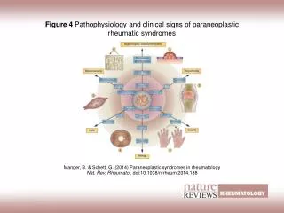
Paraneoplastic Syndromes The remote effects of cancer on the body are collectively known as paraneoplastic syndromes The visual system, the CNS, the peripheral nervous system, the neuromuscular junction and the skeletal muscles may be affected indirectly by malignancy Clinical signs may predate discovery of a primary tumor
Anti-Ri autoantibody is similar to anti-Hu It reacts with an antigen present in the nuclei of neurons throughout the central nervous system but not in glial nuclei or systemic tissues It is not species restricted in its distribution; and It is also detected in the cytoplasm of neurons but cytoplasmic staining is less prominent than the reaction with nuclei
References Budde-Steffen C, Anderson NE, Rosenblum MK, Graus F, Ford D, Synek BJL, Wray SH, Posner JB. An anti-neuronal autoantibody in paraneoplastic opsoclonus. Ann Neuron 1988; 23:528-531. Helmchen C, rambold H, Sprenger A, Erdmann C, Binkofski F. Cerebellar activation in opsoclonus: An fMRI study. Neurology 2003; 61:412-415.
Luque AF, Furneaux HM, Ferziger R, Rosenblum MK, Wray SH, Schoid SC Jr, Glantz MJ, Jaeckie KA, Biran H, Lesser MK, Paulsen WA, River ME, Posner JH. Anti-Ri: an antibody associated with paraneoplastic opsoclonus and breast cancer. Ann Neurol 1991; 29:241-251. Wong AM, Musallam S, Tomlinson Rd, Shannon P, Sharpe JA. Opsoclonus in three dimensions: oculographic, neuropathologic and modeling correlates. J Neurol Sci 2001; 189:71-81.