Chapter 25: The Digestive system
400 likes | 532 Views
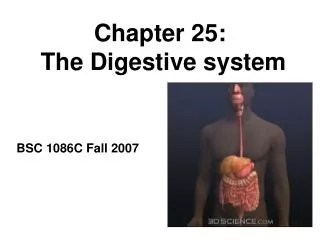
Chapter 25: The Digestive system. BSC 1086C Fall 2007. 4 basic phases of digestion. _______________________- intake of food _______________- breakdown of molecules mechanical – teeth and churning action of stomach and intestines

Chapter 25: The Digestive system
E N D
Presentation Transcript
Chapter 25: The Digestive system BSC 1086C Fall 2007
4 basic phases of digestion • _______________________- intake of food • _______________- breakdown of molecules • mechanical – teeth and churning action of stomach and intestines • chemical – hydrolysis reactions that break down macromolecules into monomers • ________________- uptake nutrients into blood/lymph • _______________- elimination of undigested material
Subdivisions of Digestive System • Digestive tract (GI tract) • 30 foot long tube extending from mouth to anus • Accessory organs • teeth, tongue, liver, gallbladder, pancreas, salivary glands 25.1
Ingestion via Mouth (Oral Cavity) • Food enters the GI tract (ingestion) via the mouth • Mechanical digestion starts as the teeth physically break down the food (increased surface area exposed to digestive enzymes!) • Chemical digestion starts due to enzymes in saliva 25.4
Salivary Glands • Small __________________ found under mucous membrane of mouth, lips, cheeks and tongue - secrete at constant rate • 3 pairs _________________ connected to oral cavity by ducts • parotid, submandibular and sublingual Secrete Saliva! 25.9
Saliva • Functions of saliva • moisten, begin starch and fat digestion, cleanse teeth, inhibit bacteria, bind food together into bolus • Hypotonic solution of 99.5% water and solutes • salivary amylase, begins starch digestion • lingual lipase, digests fat activated by stomach acid • mucus, aids in swallowing • lysozyme, enzyme kills bacteria • immunoglobulin A, inhibits bacterial growth • pH of 6.8 to 7.0 (relatively neutral) • Produce 1-1.5 L per day • Saliva is released following signals from the CNS
2: Pharynx • Skeletal muscle • deep layer – longitudinal orientation • superficial layer – circular orientation • superior, middle and inferior pharyngeal constrictors • Drive food downwards (towards esophagus) during swallowing
3: Esophagus • Straight muscular tube 25-30 cm long • esophageal glands in submucosa (lubrication) • skeletal muscle in upper part and smooth in bottom • Extends from pharynx to cardiac stomach passing through esophageal hiatus in diaphragm • Lower esophageal sphincter closes orifice to reflux (thus preventing stomach acid from entering the lower end of the esophagus causing heartburn)
4: Stomach • Mechanically breaks up food, liquifies food and begins chemical digestion of protein and fat • resulting soupy mixture is called ____________ • Does not absorb significant amount of nutrients • absorbs aspirin and some lipid-soluble drugs (alcohol)
Gross Anatomy of Stomach 25.12 • Notice: bulge of fundus, narrowing of pyloric region, thickness of pyloric sphincter and greater and lesser curvatures
Unique Features of Stomach Wall 25.13 • Mucosa • simple columnar glandular epithelium • lamina propria is filled with tubular glands (gastric pits) • Muscularis externa has 3 layers • outer longitudinal, middle circular and inner oblique layers
Cells of Gastric Glands • _____________ cells secrete mucus • __________________ cells • divide rapidly to produce new cells that migrate to surface • __________________ cells • secrete HCl acid and intrinsic factor • __________________ cells • secrete pepsinogen • chymosin and lipase in infancy • __________________ cells • secrete hormones and paracrine messengers 25.13
GastricSecretions • Parietal cells contain __________________ (CAH) CAH • CO2 + H2O H2CO3 HCO3- + H+ • H+ is pumped into stomach lumen by H+-K+ ATPase • antiporter uses ATP to pump H+ out and K+ in • HCO3- exchanged for Cl- (chloride shift) • Cl- pumped out to join H+ forming HCl 25.14 • 2 to 3 L of gastric juice/day (H2O, HCl and pepsin)
Functions of Hydrochloric Acid • Activates _________ and _______________ • Breaks up connective tissues and plant cell walls • liquefies food to form chyme • Converts ingested ferric ions (Fe3+) to ferrous ions (Fe2+) • absorbed and used for hemoglobin synthesis • Destroys ingested bacteria and pathogens
Intrinsic Factor • Intrinsic factor • A glycoprotein released by parietal cells • essential for vitamin B12 absorption by small intestine • necessary for hemoglobin production (lack causes pernicious anemia)
First: the liver Accessory digestive organs: Liver, Gallbladder and Pancreas • All release important secretions into small intestine to continue digestion
Gross Anatomy of Liver • 3 lb. organ located inferior to the diaphragm • 4 lobes - right, left, quadrate and caudate • falciform ligament separates left and right • round ligament, remnant of umbilical vein • Gallbladder adheres to ventral surface between right and quadrate lobes 25.19
Microscopic Anatomy of Liver 25.20 • Tiny cylinders called hepatic lobules (2mm by 1mm) • Central vein surrounded by sheets of hepatocyte cells separated by sinusoids lined with fenestrated_____________ • Blood filtered by hepatocytes on way to central vein
Gallbladder and Bile • Sac on underside of liver -- 10 cm long • 500 to 1000 mL bile are secreted daily from liver • Gallbladder stores and concentrates bile • bile backs up into gallbladder from a filled bile duct • between meals, bile is concentrated by factor of 20 • Yellow-green fluid containing minerals, bile acids, cholesterol, bile pigments and phospholipids • bilirubin pigment from hemoglobin breakdown • bile acid (salts) emulsify fats and aid in their digestion
Ducts of Gallbladder,Liver, Pancreas • Bile passes from _______________between cells to ______________________to right and left__________________________ • Right and left ducts join outside liver to form_________________________ • __________________from gallbladder joins common hepatic duct to form bile duct • Duct of pancreas and bile duct combine to form ________________________emptying into duodenum at major duodenal papilla 25.21
Gross Anatomy of Pancreas • Retroperitoneal gland posterior to stomach • head, body and tail • Endocrine and exocrine gland • secretes insulin and glucagon into the blood • secretes 1500 mL pancreatic juice into duodenum • water, enzymes, zymogens, and sodium bicarbonate • Pancreatic duct runs length of gland to open at sphincter of Oddi • accessory duct opens independently on duodenum 25.21
Activation of Zymogens 25.23 • _______________converted to _____________ by intestinal epithelium • Trypsin converts other 2 (also digests dietary protein)
Small Intestine 25.24 • Nearly all chemical digestion and nutrient absorption occurs in small intestine
Small Intestine • ____________curves around head of pancreas (10 in.) • retroperitoneal along with pancreas • receives stomach contents, pancreatic juice and bile • neutralizes stomach acids, emulsifies fats, pepsin inactivated by pH increase, pancreatic enzymes • ______________- next 8 ft. (in upper abdomen) • has large tall circular folds; walls are thick, muscular • most digestion and nutrient absorption occur here • ______________ - last 12 ft. (in lower abdomen) • has peyer’s patches – clusters of lymphatic nodules • ends at ileocecal junction with large intestine
Small Intestine - Surface Area • _______________(plicae circularis) up to 10 mm tall • involve only mucosa and submucosa • chyme flows in spiral path causing more contact 25.25 • ____________ are fingerlike projections 1 mm tall • contain blood vessels and lymphatics (lacteal) • nutrient absorption • ____________:1 micron tall; cover surface • brush border on cells • brush border enzymes for final stages of digestion
Intestinal Crypts • Pores opening between villi lead to intestinal crypts • absorptive cells, goblet cells and at base, rapidly dividing cells • paneth cells – antibacterial secretions • Brunner’s glands in submucosa secrete bicarbonate mucus • Peyer patches are populations of lymphocytes to fight pathogens • Secrete 1-2 L of intestinal juice/day • water and mucus, pH 7.4-7.8 25.25
Carbohydrate Digestion - Small Intestine 25.27 • Salivary amylase stops working in stomach (pH < 4.5) • 50% of dietary starch digested before it reaches small intestine • Pancreatic amylase completes first step in 10 minutes • Brush border enzymes act upon oligosaccharides, maltose, sucrose, lactose and fructose • lactose indigestible after age 4 in most humans (lactase declines)
Carbohydrate Absorption 25.28 • Sodium-glucose transport proteins (SGLT) in membrane help absorb glucose and galactose • Fructose absorbed by facilitated diffusion then converted to glucose inside the cell
Protein Digestion and Absorption 25.29 • Pepsin has optimal pH of 1.5 to 3.5 -- inactivated when passes into duodenum and mixes with alkaline pancreatic juice (pH 8)
Protein Digestion and Absorption 25.29 • Pancreatic enzymes take over protein digestion by hydrolyzing polypeptides into shorter oligopeptides • Trypsin and chymotrypsin: break peptides via hydrolysis • Carboxypeptidase: removes 1 amino acid at a time from the end of the oligopeptide
Protein Digestion and Absorption 25.29 • _____________________________finish task, producing amino acids that are absorbed into intestinal epithelial cells • ________________________, ____________________, and _________________________ • amino acid cotransporters move into epithelial cells and facilitated diffusion moves amino acids out into blood stream
Fat Digestion and Absorption 25.30 Step 1: emulsification of large fat globules by bile acids and lecithin (a phosholipid component in bile)
Fat Digestion and Absorption Step 2: _____________in the fat droplet are hydrolyzed by pancreatic lipase to form _______________________and ___________________________
Fat Digestion and Absorption 25.30 Step 3: micelles produced in the bile pick up lipids from the lumen of the small intestine
Fat Digestion and Absorption 25.30 Step 4: micelles transport lipids to brush border where they are absorbed by the intestinal cells. Complex lipids are resynthesized, packaged (chylomicron), exocytotically released and finally enter lacteal
Function of Large Intestine 25.31 • Reabsorbs water and electrolytes (NaCl) • Reduces undigested food to feces • feces = 75% water, 25% solids • solids = 30% bacteria, 30% fiber, 10-20% fat, small amounts of protein, dead epithelial cells, etc…
Anatomy of Anal Canal 25.31 • Anal canal is 3 cm total length • Anal columns are longitudinal ridges separated by mucus secreting anal sinuses
Neural Controlof Defecation 1. Filling of the rectum - stretches walls to stimulate receptors 2. Reflex contraction of rectum and relaxation of internal anal sphincter 3. Voluntary relaxation of external sphincter 25.32