Download
1 / 66
670 likes | 856 Views
Bovine Tuberculosi s Monaya Ekgatat NIAH. TB. Bovine Tuberculosis. - Introduction - Etiology & Epidemiology - Clinical Signs - Post mortem lesions - Diagnosis - Public Health - Prevention and control. NIAH. Introduction.

E N D
Bovine Tuberculosis Monaya Ekgatat NIAH TB
Bovine Tuberculosis - Introduction - Etiology & Epidemiology - Clinical Signs - Post mortem lesions - Diagnosis - Public Health - Prevention and control NIAH
Introduction Control relies on - early diagnosis - removal of infected animal - tracing - exposing infected cases Zoonosis human - aerosol - ingestion Developed countries - reduced prevalence Less developed countries - still common - economic loss NIAH
Etiology and epidemiology Agents Mycobacterium tuberculosis (human) Mycobacterium bovis (animal) Mycobacterium avium (bird) NIAH
Etiology and epidemiology Mycobacterium tuberculosis complex (MTB. Complex) M. tuberculosis M. caprae M. bovis M. pinnipedii M. africanum M. canetti M. microti
Mycobacterium bovis • Can survive for several months in the environment (cold, dark and moist condition) • 12-24 oC survival time 18-332 days • Dry or moist soil (34 oC) : 4-8 weeks • Summer : 4 days
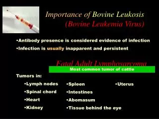
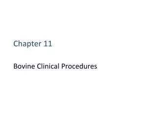
Etiology and epidemiology Maintenance hosts for M. bovis - cattle - buffalo Reservoir hosts - brush-tail possum (New Zealand) - badger (United Kingdom, Ireland) - deer (United States) - bison (Canada) - greater kudu, common duiker African buffalo, warthogs (Africa) NIAH
African buffalo Warthog
Common duiker Greater kudu
Etiology and epidemiology Transmission Respiration----aerosal (short distance) Ingestion ( unpasteurized milk) Source of infectious bacteria - respiratory secretion - feces - milk - (urine) - vaginal secretion - semen NIAH
Incubation Period • 3 weeks – years: under natural condition Morbidity and Mortality • 1-2 animals = 0 - 40% infected = 0 - 10%developed gross lesions • Severity - dose of agents - individual immunity • Mortality : rare
Clinical Signs Chronic ( rare: acute & rapidly progressive) Early infection ---- asymptomatic Late stage: symptomatic - progressive emaciation - fluctuating fever - weakness - inappetite - moist cough (pulmonary involve) - dyspnea No specific signs Asymptomatic and anergiccarriers (ill: stress, old age) NIAH
Post mortem lesions NIAH
Tubercles in liver NIAH
Diagnosis • Clinical signs (lacking) • Laboratory diagnosis • 1. Identification of the agent • a) Microscopic examination • b) Culture of M. bovis (3-6 weeks) • - biochemical tests • - culture characteristics • c) Nucleic acid recognition methods NIAH
Diagnosis 2. Delayed hypersensitivity test Tuberculin test : SID, SCITT 3. Blood-based laboratory tests a) Gamma-interferon assay b) Lymphocyte proliferation assay c) ELISA (late stages of infection, anergic cattle)
Presumptive Diagnosis • Histopathology • Microscopic demonstration of acid-fast bacilli • Direct smear from clinical samples/tissues and stained with Z-N stain, fluorescent acid-fast stain or immunoperoxidase
Differential Diagnosis - Contagious bovine pleuropneumonia -Pasteurella or Corynebacterium pyogenes pneumonia - Aspiration pneumonia ( secondary infection) - Traumatic pericarditis - Caseouslymphadenitisor melioidosis (small ruminant) - Chronic aberrant liver fluke infestation NIAH
Laboratory Diagnosis 1. Identification and isolation of the agent NIAH
Immunohistochemistry NIAH
DNA - hybridization NIAH
ELISA: Detection of MTB complex 1 2 3 1 2 3 1 2 3 1 2 3 NIAH
The immune system : Antigen Antigen presenting cells (Macrophages & reticulocites) Humoral immunity Cell mediated immunity Memory B-lymphocytes T-lymphocytes Plasma cells antibodies Lymphokines cytooxicity Diagnostic measures of an Immune response Skin test / Lymphocyte stimulation / Gamma-interferon ELISA
2.Tuberculin test Antigen:Bov. PPD 0.1 ml(not more than 0.2 ml)= 2,000 IU – 5,000 IU Work plan 0h 244872hrs injection 1stmeasure 2ndmeasure Negative reaction < 2 mm w/o local clinical signs Inconclusive reaction 2-4 mm w/o local clinical signs Positive reaction ≥ 4 mm with or w/o local clinical signs (one fold + ≥ 8 mm) Retest: after 42-60 days (cattle), 120 days (deer) NIAH
Tuberculin test NIAH
Positive reaction NIAH