Understanding Antibodies: Structure, Types, and Functions in Immune Responses
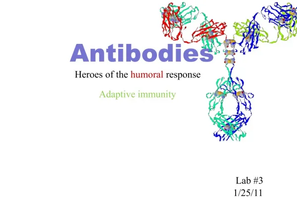
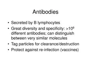
Antibodies, also known as immunoglobulins, are proteins secreted by plasma cells that bind specifically to antigens. They originate from B cells and come in various forms depending on the epitopes they target. Antibodies consist of four peptide chains: two identical light chains and two identical heavy chains, linked by disulfide bonds. Their variable regions allow for a diverse range of antigen binding sites, while constant regions determine their class (e.g., IgG, IgA). Essential for immune function, antibodies are involved in opsonization, complement activation, and transcytosis at mucosal surfaces.

Understanding Antibodies: Structure, Types, and Functions in Immune Responses
E N D
Presentation Transcript
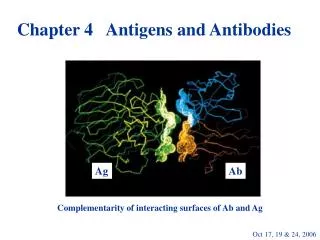
Chapter 4 Antibodies • Ab’s are Ag-binding proteins secreted by plasma cells; found on surfaces of B cells and free in the blood/IF/lymph • Ab’s triggered by an Ag are heterogeneous -produced for dif’t epitopes on the Ag (polyclonal Ab’s) -from several clones of B cells – each producing monoclonal Ab’s
Antibody isolation • Centrifuged blood contains: • Cells • Liquid (plasma) • If left to clot – serum • Serum is Ab rich • Electrophoretic peaks shown by Tiselius and Kabat (1939) Injection of ovalbumin
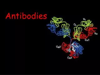
Antibody structure • 4 peptide chains: • 2 identical Light chains • 2 identical Heavy chains • Ea. light chain is bound to a heavy chain by di-S bond + noncovalent attractions • Similar di-S bonds link 2 H-L chain combo’s (H-L)2
Antibody structure • 1st 110 aa’s @ NH3 end exhibit variation in both H + L chains -(V)ariable regions • w/i V regions are CDR’s – specific binding sites for Ag • w/i the same Ab class, there is relatively constant aa seq thru rest of molecule – (C)onstant regions • Glycolsylation of Fc fragments – Ab’s are glycoproteins
Discovery of aa sequences in H and L chains • Each Ag carries multiple epitopes (even haptens!) so that initial efforts hindered • Discovery of multiple myeloma (and Bence Jones proteins) allowed isolation of large quant. of homogenous Ab • Can stimulate MOPC’s in mice from intra-peritoneal mineral oil injections stim production of malignant plasma cells
Light Chain sequencing: • V regions (amino terminal half) vary in aa sequence • C regions (carboxyl terminal half) show 2 basic aa sequence patterns • Led to typing of L chains – kappa (κ) and lambda (λ) • minor differences in λ chains results in further sub-typing ( λ1, λ2, and λ3)
Heavy Chain sequencing: • Like L chains, amino terminal end reveals variation in 1st 100-110 aa’s • Remaining portion revealed 5 seq. patterns corresponding to 5 dif’t C regions: labelled γ, α, μ, δ, and ε • Each of these 5 C chains creates an Isotype • Heavy chains of Ab’s determine the class of the Ab – IgG, IgA, IgM, IgD, IgE • Minor differences in α and γ chains (α1,2 and γ1,2,3,4) called “subisotypes”
Diversity of Variable Regions is concentrated in CDR’s • Max variation is seen in those aa sequences in loops joining β sheets of the proteins of the VL and VH regions Ag binding sites • As Ag-binding occurs between complementary aa’s = “complementarity-determining regions” (CDR’s) • there are 3 such loops on both chains • Variation in length and sequence of the 6 CDR’s creates wide range of Ab specificities!!
Constant region domains They perform a variety of functions: • CH and CL domains support and orient (V)ariable regions contrib to > diversity • “Hinge” region between CH1 and CH2 rich in proline; di-S bonds between cysteines • IgG, D, A have 3 C domains (CH1-3) • IgM, E have 4 C domains (CH1-4) • CH2’s of G,D,A and CH3’s of M, E are separated by polysaccharides (solubility) and Complement binding sites • CH3’s and CH4’s exhibit carboxyl-terminal ends • Secretory Ig has hydrophilic aa’s @ COOH end • Membrane Ig contains 3 parts:
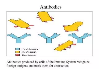
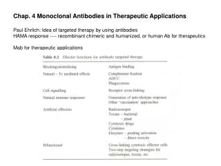
Roles of Antibody: Both ends of Ab are functional (NH3-end: Fab; COOH-end: membrane + protein binding) • Opsonization • Complement activation • Transcytosis to mucosal surfaces