Download

1 / 179
1.81k likes | 2.44k Views
Chapter 15 Diseases Resulting from Fungi and Yeasts. Andrews’ Diseases of the Skin JoAnne M. LaRow, D.O. Superficial mycoses. AKA dermatophytes Classified into three genera: Microsporum, Trichophyton, Epidermophyton

E N D
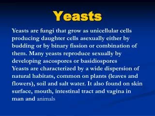
Chapter 15Diseases Resulting from Fungi and Yeasts Andrews’ Diseases of the Skin JoAnne M. LaRow, D.O.
Superficial mycoses • AKA dermatophytes • Classified into three genera: Microsporum, Trichophyton, Epidermophyton • Mycoses caused by dermatophytes are called dermatophytosis, tinea, ringworm • On certain parts of body tinea has certain features characteristic of that site • Hence the division into seven types (1)tinea capitis, (2)tinea barbae, (3)tinea faciei, (4)tinea corporis, (5) tinea manus, (6) tinea pedis, (7) tinea cruris, (8)onychomycosis • Superficial mycoses can be divided into causative dermatophyte- • Management is rarely assisted by ID of genus and species
Susceptibility • They are soil saprophytes that have acquired ability to digest keratinous material in soil, becoming “keratinophilic fungi” • Some have evolved to parasitize keratinous tissues of animals frequently in contact with soil and have lost their ability to survive in soil (zoophilic fungi) • Anthropophilic dermatophytes are believed to have evolved from zoophilic fungi, adapting to human keratin and losing their ability to digest animal keratin • Environmental conditions help promote propagation of many opportunistic fungi • Host factors are also significant
Host factors • Immunosuppressed pts • Pts with AIDS may have severe forms • Genetic susceptibility to certain forms of fungal infections may be related to types of keratin or degree or mix of cutaneous lipids produced • Surface antigens-ABO system-one study of 108 culture proven dermatophytosis pts noted that pts with type A blood were prone to chronic disease • Human steroid hormones can inhibit growth of dermatophytes (androgens like androstenedione) • One group believes this high susceptibility of Trichophyton rubrum & Epidermophyton floccosum to intrafollicular androstenedione is a reason why these species do not cause tinea capitis
Antifungal therapy • Consider spectrum of activity of antifungal • Pharmacokinetic profile of the agent • Clinical type of infection • Additionally, safety, compliance and cost • Griseofulvin is still therapeutic option but studies are showing that newer antifungals are more efficacious
Imidazoles • Clotrimazole, miconazole, sulconazole, oxiconazole, and ketoconazole • Mostly used for topical tx • Inhibit cytochrome P450 14-alpha-demethylase (an essential enzyme in ergosterol synthesis) • Ketaconazole has wide spectrum against dermatophytes, yeasts, and some systemic mycoses • Ketaconazole has the potential for serious drug interactions and a higher incidence of hepatotoxicity during long-term daily therapy
Allylamines • Naftifine, terbinafine, butenafine • Mode of action similar to thiocarbamates • Inhibites squalene epoxydation • Terbinafine has less activity against Candida species in vitro studies then triazoles, but is effective clinically • Terbinafine is ineffective in the oral tx of tinea versicolor but is effective topically • Few drug interactions have been reported, bioavailability is unchanged in food, hepatotoxicity, leukopenia, severe exanthems, and taste disturbances occur uncommonly but should be monitored for clinically and by lab testing if continuous dosing over 6 weeks occurs
Polyene • Nystatin • Irreversibly binding to ergosterol-an essential component of fungal cell membranes
Triazoles • Itraconazole • Fluconazole • Affect P450 system • Numerous drug interactions occur • Need to know pt’s current meds • Broadest spectrum to dermatophytes and Candida species, and Malassezia furfur • Itraconazole is fungistatic-food increases its absorption , antacids and gastric acid secretion suppressors produce erratic or lowered absorption • Pulse dosing limits concern over lab abnormalities • Fluconazoles’s absorption is unaffected by food
Tinea Capitis • Occurs chiefly in children – less commonly in infants and adults • Boys more frequently than girls; except in epidemics caused by Trichophyton tonsurans where there is equal frequency • Divided into inflammatory and noninflammatory • Tinea capitis can be caused by all pathogenic dermatophytes except Epidermophyton floccosum and T. concentricum • In U.S. most caused by T. tonsurans(replacing Microsporum audouinii) & M. canis
Noninflammatory • M. audouinii infections present as the classic form • Characterized by multiple scaly lesions (“gray-patch”), stubs of broken hair, and a minimal inflammatory response • Occasionally glabrous skin, eyelids, and eyelashes are involved • Sometimes observed in epidemics in schools and orphanages • Over past 30 yrs, M. audouinii infections are being replaced by increasing numbers of “black-dot” ringworm, caused primarily by T. tonsurans and occassionally by T. violaceum • In the U.S. T. tonsurans is the most common cause
Tinea Capitis • “Black dot” ringworm, caused by T. tonsurans & occasionally T. violaceum presents as multiple areas of alopecia studded with black dots representing infected hairs broken off at or below the surface of the scalp
Inflammatory • Usually caused by M. canis • Can be caused by T. mentagrophytes, T. tonsurans, M. gypsem, or T. verrucosum • M. canis infection begins as scaly, erythematous, papular eruptions with loose and broken-off hairs, followed by various degrees of inflammation • A localized spot accompanied by pronounced swelling, with developing bogginess and induration exuding pus develops-kerion celsii • A delayed type hypersensitivity reaction to fungal elements • With extensive lesions fever, pain, and regional lymphadenopathy may occur
Kerion • Widespread “id” eruptions may appear concomitantly on trunk and extremities • These are vesicular, lichenoid, or pustular • Kerion may be followed by scarring and permanent alopecia in areas of inflammation and suppuration • Systemic steroids for short periods will greatly diminish the inflammatory response and reduce the risk of scarring
Kerion: inflammatory rxn of tinea capitis caused by Microsporum canis or Trichophyton mentagrophytes
Favus • Rare in the U.S. • Appears mainly on the scalp, but may occur on glabrous skin and nails • On scalp, concave sulfur-yellow crusts from around loose, wiry hairs • On glabrous skin lesions are pinhead to 2 cm in diameter with cup-shaped crusts called scutulae-usually pierced by a hair as on the scalp • Scutula have a distinctive mousy odor • Nail involvement causes brittle, irregularly thickened, and crusted nail changes • Not seen typically in North America(has been reported in Kentucky and Canada) • Called witkop in South Africa by the Bantus
Etiology • Tinea capitis can be cause by any one of several species: T. tonsurans, M. audouinii, and M. canis • First two are spread from human to human • Latter is caught from animals such as kittens and dogs • Most frequent invaders of scalp are endothrix types-T. tonsurans(black-dot ringworm) and T. violaceum • T. tonsurans alone affects adults(chiefly women) regularly; others affect children • Ectothrix found on scalp are T. verrucosum & T. mentagrophytes (less frequently seen is T. megninii-southwest Europe)
Pathogenesis • Incubation period lasts 2 to 4 days • Hyphae grow downward into the follicle, on the hair’s surface, and the intrafollicular hyphae break up into chains of spores • Period of spread (4 days to 4 months) during which lesions enlarge and new lesions appear • At about 3 weeks hairs break off a few millimeters above the surface • Intrapilary hyphae descend to exact upper limit of keratogenous zone and here form Adamson’s “fringe” on the twelfth day • External portions of intrapilary hyphae segment into chains of ectothrix spores
Pathogenesis • No new lesions develop during the refractory period (4 months to several yrs) • Clinical appearance is constant-with host and parasite in equilibrium • This is followed by a period of involution in which the formation of ectothrix spores and intrapilary hyphae gradually diminishes • Asymptomatic carrier states among young black children may occur • There has been a lack of correlation between number of asymptomatic carriers and index cases-suggesting that carrier cases are not primary mode of transmission of T. tonsurans
Histology • Extensive inflammation leading to follicular destruction
Medium power: dense inflammation consists of mixed cell types
Neutrophils and other inflammatory cells surround this small follicle. • Fungal elements are present within hair shaft
Diagnosis • Ultraviolet of 365 nm wavelength is obtained by passing a beam through a Wood’s filter composed of nickel oxide-containing glass • This apparatus a Wood’s light , is available commercially • A simple form is the 125-volt purple bulb • In a dark room the skin under this light fluoresces faintly blue; however, infected hairs fluoresces bright green, beads on the hairs contrasting strongly with the dark field • Bare, scaly areas show a turquoise blue color • Fluorescent-positive infections are caused by :M. audouinii, M. canis, M. ferrugineum, M. distortum, T. schoenleinii
Diagnosis • Hairs infected with T. tonsurans & T. violaceum and others of endothrix do not fluoresce • The fluorescent substance is pteridine • For microscopic demonstration of the fungus, two or three loose hairs are removed • Hairs are placed on slide with a drop of 10-20% solution of KOH • A cover slip is applied, specimen is warmed until hairs are macerated • Examine under low, then high power • Xylol is as satisfactory as KOH and need not be warmed • Scales or hairs cleared with it can still be cultured
Diagnosis • Fungus invades hair shaft in two ways-(1) ectothrix involvement in which hair is surrounded with a sheath of tiny spores • Examples of these types are: Microsporum species, T. mentagrophytes & T. verrucosum (T. verrucosum is the fungus most frequently acquired by humans from cattle and causes a severe inflammatory tinea barbae in men or tinea capitis in children) • Other mode of infection is endothrix type-where arthrospores are formed inside the hair shaft • This type is seen in T. tonsurans, T. violaceum, and T. schoenleinii infections
Final and exact identification of causative fungus • Such identification is largely epidemiologic and academic-tx is the same • Several infected hairs are placed on Sabouraud’s glucose agar or Dermatophyte Test Medium (DTM) • On DTM a distinctive growth appears within 1-2 weeks • Diagnosis is usually made by gross appearance of culture • When questionable the culture is examined under a microscope for characteristic morphologic forms • DTM contains antibiotics to reduce growth of contaminants and a colored pH indicator to denote the alkali-producing dermatophytes
DTM • A few nonpathogenic saprophytes will also produce alkalinization and in the occasional case of onychomycosis of toenails caused by airborne molds, a culture medium containing an antibiotic may inhibit growth of the true pathogen • Cultures are best taken by rubbing the lesion vigorously with a sterile cotton swab moistened with sterile water and them streaked over the agar surface
Ectothrix type in Microsporum canis-note small spores around hair shaft
Endothrix in T. scoenleinii showing characteristic bubbles of air
Endothrix infection, (low-power KOH mount): arthroconidia noted within hair shaft • Endothrix infection (high-power KOH mount) showing total hair shaft involvement
T. tonsurans • This microoraganism grows slowly in culture to produce a granular or powdery yellow to red, brown, or buff colony • Crater formation with radial grooves may be produced • Microconidia may be seen regularly • Dx confirmed by the fact that cultures grow poorly or not at all without thiamine
T. mentagrophytes • Cultural growth is velvety or granular or fluffy, flat or furrowed, light buff, white, or sometimes pink • Back of the culture can vary from buff to dark red • Round microconidia borne laterally and in clusters confirm dx within 2 weeks • Spirals are sometimes present • Macroconidia may be seen
T. verrucosum • Growth is slow and cannot be observed well for at least 3 weeks • Colony is compact, glassy, velvety, , heaped or furrowed, and usually white, but may be yellow or gray • Chlamydospores are present in early cultures • Microconidia may be seen
M. audouinii • Gross appearance shows a slowly groing, matted, velvety, light brown colony • Back of which is reddish brown to orange • Under microscope a few large multiseptate macroconidia (macroaleuriospores) are seen • Microconidia (microaleuriospores) in a lateral position on hyphae are clavate • Racquet mycelium, chlamydospores, and pectinate hyphae are seen sometimes
M. canis • Culture shows profuse, fuzzy, cottony, aerial mycelia tending to become powdery in the center • Color is buff to ligth brown • Back of colony is lemon to orange-yellow • Numerous spindle-shaped multiseptate microconidia and thick-walled macroconidia are present • Clavate microconidia ae found along with chlamydospores and pectinate bodies
Treatment • Griseofulvin of ultramicronized form, 10 mg/kg/day, is the daily dose recommended for children • Grifulvin V is the only oral suspension available for children unable to swallow tablets-dose is 20 mg/kg/day • Tx should continue for 2-4 months, or for at least 2 weeks after a negative microscopic and culture examinations are obtained • Griseofulvin does not primarily affect the delayed type hypersensitivity reaction responsible for the inflammation in kerion • For this, systemic steroids, to minimize scarring, can be given simultaneously
Numerous other studies exist that demonstrate the effectiveness of other oral agents, such as itraconazole, terbinafine, and fluconazole • These studies report these meds to be excellent alternatives, but the total reported experience to date is low • Selenium sulfide shampoo or ketaconazole shampoo three times weekly can be used as adjunctive therapy to oral antifungal agents • Herbert recommends culture of family members, caution regarding sharing potentially contaminated fomites, and simultaneous tx of all persons infected clinically or by culture • Drake et al recommend tx family members with ketaconazole shampoo, selenium sulfide shampoo, or povidine-iodine even if they are asymptomatic