Download
1 / 83
830 likes | 963 Views
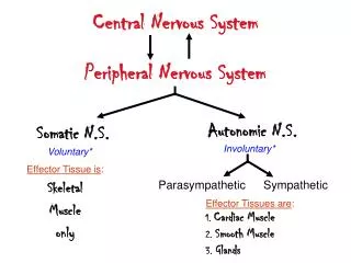
Cardiac & Nervous System Review of Emergencies. ECRN Mod III CE 2010 Condell Medical Center EMS System Prepared by: FF/PMD Michael Mounts Lake Forest Fire Department Reviewed/revised by: Dr. Kent Bailey, EMS Medical Director. Objectives. Identify components of the nervous system

E N D
Cardiac & Nervous SystemReview of Emergencies ECRN Mod III CE 2010 Condell Medical Center EMS System Prepared by: FF/PMD Michael Mounts Lake Forest Fire Department Reviewed/revised by: Dr. Kent Bailey, EMS Medical Director
Objectives • Identify components of the nervous system • Identify signs and symptoms of a patient with a CVA • Identify assessment & field treatment of patient with a CVA • Identify anatomy and physiology of the cardio-pulmonary system • Identify signs and symptoms of a patient with ACS • Identify field treatment of patient with ACS
Objectives cont. • Discuss situations for using the RAD 57 tool • Identify patient care based on RAD 57 readings • Review documentation components for discussed conditions • Identify a variety of ECG rhythm strips • Review Region X SOP’s for various emergencies discussed
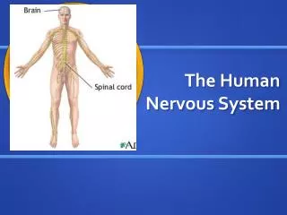
Components of the CNS • Brain - 3 major structures • Cerebrum • largest element of nervous system • occupies most of cranium • highest functional portion of brain • center of conscious thought, personality, speech, motor control, and visual, auditory, & tactile perception • Cerebellum • fine tunes motor control, allows smooth motion from one position to another • responsible for balance & maintenance of muscle tone
Components of the CNS cont. • Brainstem • central processing center &communication junction • midbrain • hypothalamus • controls much of endocrine function, vomiting reflex, hunger, thirst, kidney function, body temperature
Components of the CNS cont. • Brainstem cont. • pons • medulla oblongata • respiratory center (depth, rate, rhythm) • cardiac center (rate & strength of cardiac contractions) • vasomotor center (control of distribution of blood and maintenance of blood pressure)
Cross-section of the brain skull periosteum In order… 1. Skull bone 2. Periosteum of the skull 3. Dura 4. Arachnoid 5. Subarachnoid space 6. Pia mater dura
CNS Circulation • 4 major arterial vessels • Capillaries unique • walls thicker so they are less permeable • protected environment via the blood-brain barrier • Cerebral perfusion • changes in ICP are met with compensatory changes in blood pressure
Cerebral Perfusion Pressure • Intracranial pressure - pressure within cranium • pressures within cranium create a natural resistance to control the amount of cerebral blood flow • blood flow to the brain remains adequate as long as pressures within the cranium are appropriate • 3 major cranial contents • brain, blood, & cerebrospinal fluid • Any changes in one of the 3 cranial contents is at the sacrifice to one of the others • When ICP rises, the body increases the BP to maintain the cerebral perfusion (Cushing reflex)
Brain Function By Region • Frontal Lobe - reasoning, planning, parts of speech, movement, emotions, and problem solving • Parietal Lobe - movement, orientation, recognition, perception of stimuli • Occipital Lobe - visual processing • Temporal Lobe - perception and recognition of auditory stimuli, memory, and speech • Cerebellum - regulation and coordination of movement, posture, and balance • Brain stem - breathing, heartbeat, and blood pressure
Remember • Wernicke’s Area • Controls speech comprehension • Broca’s Area • Controls speech production • Both on left side of brain • If either of the above speech areas are noted to be affected, see if right sided weakness is also present • Speech and motor problems will be reflected on opposite sides of the body
Left vs. Right • This theory of the structure and functions of the mind suggests that the two different sides of the brain control two different “modes” of thinking. It also suggests that each of us prefers one mode over the other. Left Brain Logical Sequential Rational Analytical Objective Looks at parts Right Brain Random Intuitive HolisticSynthesizing Subjective Looks at wholes
Left vs. Right cont. Note: Notice how Broca & Wernicke’s area are on Left side Hearing difference: Speech on Left vs. Music on Right
CVA Signs and Symptoms • Trouble with walking, sudden dizziness, loss of balance or loss of coordination. • Trouble with speaking and/or understanding, confusion, slurred words or be unable to find the right words to explain what is happening (aphasia). • Paralysis or numbness on one side of the body or face. • Trouble with seeing in one or both eyes. Sudden blurred or blackened vision, or seeing double. • Headache; a sudden, severe "bolt out of the blue" headache which may be accompanied by vomiting, dizziness or altered consciousness.
What to do… • Initial assessment • AVPU, ABC’s, life threats, etc. • Sample history • Vitals • Pupils • Glasgow • Time of onset VERY important! • F.A.S.T. or Cincinnati Stroke Scale • Remember… you only need to have one of these signs for positive CVA identification.
Cincinnati Stroke Scale or FAST • F – look for facial drooping • Have patient smile large enough to see teeth • A – check for arm drift • Patient holds hands out in front for 10 seconds with eyes closed, palms up • S – check for slurred speech • T – teach patients to call 911 – time is essential
Facial Drooping • Ask the patient to smile real big and show you their teeth • Best way to see if a droop is present
Arm Drift • Demonstrate first and then have patient hold their hands out in front, palms up, for 10 seconds
Clarity of Speech • Most likely you’ll know by now if there is a speech problem • Can have the patient repeat after you any words or a sentence you give them • “You can’t teach an old dog new tricks”
7 D’S Of Stroke Care • Detection – of signs and symptoms • Dispatch – advise to call 911 • Delivery – to the appropriate facility • Door – emergent triage in the ED • Data – appropriate tests • Decision – to administer a fibrinolytic or not • Drug – must administer the fibrinolytic within 3 hours of onset of symptoms
Intracranial Hemorrhages • Epidural – rapid onset, traumatic • Arterial bleed • Headache • Nausea/vomiting • Seizures • Focal neurologic deficits (aphasia, weakness, numbness) • Subdural – slower onset, traumatic • Venous bleed • Symptoms are often vague • Usually altered mental status • Seen more often in elderly; brain atrophy stretches the veins, making them more likely to tear in trauma *Note - White area is bleeding
Intracranial Hemorrhages • Subarachnoid – sudden onset • Usually from berry aneurysm rupture from the base of the brain; bleeding around the brain (mixed with the CSF) • Usual spontaneous, non-traumatic • Sudden severe headache • Vertigo • Light sensitivity • Often altered mental status • Intraparenchymal (inside brain tissue) • Traumatic bleed or spontaneous rupture of AVM (arteriovenous malformation)
Cardio-Pulmonary A&P • We need to know what is being affected and how that is shown as sign and/or symptoms • Knowing the following general A&P will assist in assessment • Veins • Arteries • Other tissues
Coronary Circulation • Coronary arteries and veins • Myocardium extracts the largest amount of oxygen as blood moves into general circulation • Oxygen uptake by the myocardium can only improve by increasing blood flow through the coronary arteries • If the coronary arteries are blocked, they must be reopened if circulation is going to be restored to that area of tissue supplied
The Electrical Conduction System • SA Node • AV Node • Bundle of HIS • Purkinje Fibers
The Electrical Conduction System cont. • SA node: Fastest rate of automaticity automaticity. “Primary” pacemaker of the heart. Rate: 60 to 100 bpm • AV node: Has a delay which allows for atrial contraction and a more filling of the ventricles. Rate: 40-60 bpm (if not driven by the rate above) • Bundle of His: Has the ability to self-initiate electrical activity Rate: 40-60 bpm • Purkinje Fibers: Network of fibers that carry electrical impulses directly to ventricular muscle. Rate: 20-40 bpm (if not driven by the rate above)
Electrocardiogram (ECG/EKG) • It’s name is made of 3 different parts: • electro, because it is related to electrical activity • cardio, Greek for heart • gram, a Greek root meaning "to write"
12-Lead Electrodes • A lead is a tracing of the electrical activity between 2 electrodes • Leads view the heart from the front of the body • Top, bottom, right, and left side of heart • Leads view the heart as if it were sliced in half horizontally • Front, back, right, and left sides of heart • Each lead has a positive and a negative electrode
12-lead ECG • A 12-lead ECG is made up of a tracing of the electrical activity of the heart from 12 different points of view. The point of view comes from the location of the positive electrode of each lead. The positioning of these electrodes is broken down into 3 categories; • The limb leads (lead I, II & III) • The augmented leads (aVR, aVL & aVF) • The precordial/chest leads (V1, V2, V3, V4, V5,V6)
Standard 12-Lead EKG • Six limb leads • Leads I, II, III, aVR, aVL, aVF • Six chest leads (precordial leads) • V1, V2, V3, V4, V5, V6 • Information from 12 leads obtained from the attachment of only 10 electrodes
Contiguous ECG Leads • EKG changes are significant when they are seen in at least two contiguous leads • Two leads are contiguous if they look at the same area of the heart or they are numerically consecutive chest leads
Lateral Wall MI: I, aVL, V5, V6 Source: The 12-Lead ECG in Acute Coronary Syndromes, MosbyJems, 2006.
Complications of Lateral Wall MI • I, aVL, V5,V6 • Complications arise due to the conduction components that are in the septum • Conduction dysrhythmias most common • Second degree Type II – classical • 3rd degree – complete heart block • Bundle branch blocks • Monitor patient closely for these blocks • 2nd degree Type II and 3rd degree are serious dysrhythmias that need to be treated aggressively with TCP
Inferior Wall MI: II, III, aVF Source: The 12-Lead ECG in Acute Coronary Syndromes, MosbyJems, 2006.
Complications of Inferior Wall MI • II, III, aVF • 40% of patients with inferior MI’s have right ventricular infarcts • In the presence of a right ventricular infarct, there is a high likeliness of both ventricles being damaged • Contraction capabilities will be negatively affected • Patients may present hypotensive • Nitrates and Morphine alone will dilate blood vessels worsening hypotension • Under Medical Control direction patients are often treated with a fluid challenge with the nitrates • 1st degree heart block and Second degree Type I Wenckebach most common heart blocks
Septal MI: V1 and V2 Source: The 12-Lead ECG in Acute Coronary Syndromes, MosbyJems, 2006.
Complications of Septal Wall MI • V1 and V2 • Significant amount of conduction components are in the septal area • Patient predisposed to dysrhythmias • Second degree Type II – classical • 3rd degree heart block • Bundle branch block • Lethal heart blocks treated aggressively - TCP • Rare to have a septal MI alone • Common to have anterior or lateral involvement along with septal area
Anterior Wall MI: V3, V4 Source: The 12-Lead ECG in Acute Coronary Syndromes, MosbyJems, 2006.
Complications of Anterior Wall MI • V3, V4 • Known as the “widowmaker” due to the potential for a massive area of infarction from blockage of the large amount of myocardium supplied by the LAD (left anterior descending artery) • Often the septal or lateral walls are also involved • Watch for lethal ventricular dysrhythmias and cardiogenic shock • Second degree Type II and 3rd degree heart block are more common than other blocks
Anterior Wall MI cont. • Early death within a few days often from CHF • Massive area of ventricular tissue infarcted if LAD totally occluded • Important to obtain history of recent MI diagnosis and hospital discharge • Increased incidence of ventricular tachycardia (VT) and ventricular fibrillation (VF) up to 1 -2 weeks post acute anterior MI
Posterior MI: Reciprocal Changes ST Depression V1, V2, V3, poss V4 Source: The 12-Lead ECG in Acute Coronary Syndromes, MosbyJems, 2006.
Atypical Presentation in the Elderly • Most frequent symptoms of acute MI: • Shortness of breath • Fatigue and weakness (“I just don’t feel well”) • Abdominal or epigastric discomfort • Often have preexisting conditions making this an already vulnerable population • Hypertension • CHF • Previous AMI • Likely to delay seeking treatment
Discomfort described as: Aching Tightness Pressure Sharpness Burning Fullness Tingling Often have no actual chest pain to offer as a complaint. Often the pain is in the back, shoulders, or neck Frequent acute symptoms: Shortness of breath Weakness Unusual fatigue Cold sweats Dizziness Nausea/vomiting Atypical Presentation in Women
Atypical Presentation in the Patient With Diabetes • Atypical presentation due to autonomic dysfunction • Common signs/symptoms: • Generalized weakness • Generalized feeling of not being well • Syncope • Lightheadedness • Change in mental status
Remember… • Watch out for the “triple threat” • Elderly • Female • Diabetic history • How many elderly women with diabetes do you see in your facility? • Probably lots!!!
Using Region X Cardiac SOP’s • Care is initiated for all patients based on physical assessment • A pediatric patient is considered under the age of 16 (15 and less) • EMS is not to delay care to contact Medical Control – call after care initiated • But, prompt communication is encouraged