Download
1 / 48
520 likes | 862 Views
Biology 9. Unit 9: The Cellular Basis of Reproduction and Inheritance. Objectives: After completing this Learning Quest the student will…. Create a step by step diagram of how genes pass from cell to cell. Describe the cell cycles and the process of mitosis and meiosis.

E N D
Biology 9 Unit 9: The Cellular Basis of Reproduction and Inheritance.
Objectives: After completing this Learning Quest the student will… • Create a step by step diagram of how genes pass from cell to cell. • Describe the cell cycles and the process of mitosis and meiosis. • Describe what happens when abnormalities occur in the meiosis. • Locate and describe alternations of chromosome structures.
Directions • Follow the instructions in the Anticipation Guide found in this PowerPoint Presentation. • Follow the instructions and answer all questions found in the Learning Guide. • Follow the instructions in the Conclusion Guide. • ALL THREE GUIDES CAN BE FOUND IN THIS LEARNING POWERPOINTQUEST.
Anticipation Guide (Page 1) • The center of the cell not only controls the cells activities but it also initiates and controls the time when your cell’s should reproduce.
Learning Guide (Page 1) • During cell reproduction, a cell will divide into two identical cells.The original cell is called a “parent” cell while the two new resulting cells are called “daughter” cells.
Learning Guide (Page 2) • Reproduction of single cell organisms occur through a process known as asexual reproduction. • Asexual reproduction does not require fertilization of an egg by a sperm.Offspring that are produced asexually can allow newly formed daughter cells to inherit their chromosomes from one parent cell.
Learning Guide (Page 3) • On the other hand, sexual reproduction does involve the fertilization of an egg by a sperm. This process involves a special type of cell division/reproduction called meiosis which only occurs in reproductive organs. .
Learning Guide (Page 4) • Before we can talk about the cell cycle and cell division let’s talk about chromosomes.
Learning Guide (Page 5) • Chromosomes are coiled up strands of DNA that contain the genetic material required by the cell. • These chromosomes are found within the nucleus of all plant and animal cells. • These chromosomes will play an important role in the development of genetic characteristics.
Learning Guide (Page 6) Chromatin • Chromosomes are long thread like structures that are only visible under the microscope. • In non-dividing cells, chromosomes are seen as a combination of DNA and protein molecules called chromatin. As seen in the diagram to the right.
Learning Guide (Page 7) • Notice that inside each chromosome is one long strand of DNA super coiled up around special proteins called histones. • The 46 human chromosomes found in each one of our cells contain an estimated 25 thousand genes.
Learning Guide (Page 8) • The passing on of genetic information from one cell to another occurs when a parent cell divides into two daughter cells. Mitosis is responsible for creating body cells that are “diploid” because they contain the full compliment of chromosomes. Humans have 46 chromosomes per cell. The cell cyclediagram to the right shows the process your body cells continually go through.
Learning Guide (Page 9) • The Cell Cycle consists of two major phases, interphase and mitosis. • Interphase is the first step of the cell cycle where the chromosomes make identical copies of themselves. This replication process creates an exact set of instructions to be passed on to the new cell. • During the mitotic phase the duplicated chromosomes will now divide evenly among the two daughter cells.
Learning Guide (Page 10) • The first step in the cell cycle is called Interphase. This is the longest phase of the cell cycle and represents a normal nondividing cell. During interphase the cell grows during the G 1 phase, makes an identical set of chromosomes during the S phase (replication) and forms an additional set of organelles during the G 2 phase.
Learning Guide (Page 11) • During the process of mitosis 4 additional steps take place. It begins with a stage called Prophase where the nucleus disappears and spindle fibers attach to the chromosomes that are now visible. • Inside the nucleus the chromatin fibers transform from a disorganized group of fibers to thick organized strands called chromosomes viewable with a light microscope.
Learning Guide (Page 12) • In the final part of prophase the nuclear membrane breaks apart.This will allow the microtubule spindles to reach out and connect to the chromosomes. • These protein fibers will help move and distribute the chromosomes.
Learning Guide (Page 13) • During Metaphase, the chromosomes are positioned along the middle of the cell. Refer to the diagram to the left for the correct placement of the chromosomes.
Learning Guide (Page 14) • During Anaphase, the chromosomes are pulled to the opposite ends of the cell. As a result, an equal amount of genetic information/chromosomes will be at each end of the cell or the”poles”.
Learning Guide (Page 15) • During Telophase, each cell will have received one half of the chromosomes. • Telophase is the direct opposite of prophase. • During Telophase, the nuclear membrane begins to reform as the chromosomes unravel. • The final stage of cell division is called cytokinesis. During this stage the cytoplasm and organelles are divided between the two daughter cells.
Learning Guide (Page 16) • The process where sexual reproduction of cells occur is called Meiosis. Meiosis is responsible for creating sex cells/gametes that contain only one half the chromosomes. These gametes therefore are “haploid” (contain one half the chromosomes) and are also genetically different from the parent cell. • During the life cycle living multicellular organism move through a stages of life leading from the adult stage of one generation to the adult stage of the next generation. Chromosomes also follow through from one generation to the next however with slight alterations between the generations.
Learning Guide (Page 17) • Humans are often known as diploid organisms because all of our cells contain two identical chromosomes. • The total number of chromosomes in humans are 46. The exception to his are the egg and sperm cells which are known as gametes.
Learning Guide (Page 18) • Gametes, made by a process called meiosis, contain a single set of chromosomes. To be exact, 22 autosomes and a single sex chromosome, either X or Y.
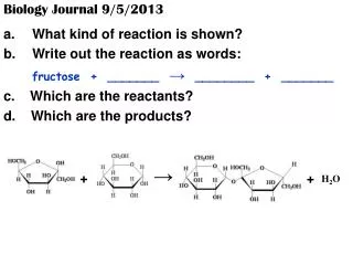
Learning Guide (Page 19) • A cell that has a single chromosome set is known as a haploid cell. • During sexual intercourse a haploid sperm cell fuses with the haploid egg cell during the process called fertilization. The results of this fertilized egg is called a zygote. This zygote now is a diploid organism containing two sets of chromosomes, one set from each parent. • The figure below demonstrates how meiosis creates chromates used in the production of chromosomes for fertilization.
Learning Guide (Page 20) • The differences between Mitosis and Meiosis can be viewed in the diagram below. As seen in the process of meiosis below the parent cell will first create two haploid cells and then in the Meiosis II the haploid cell divides again to create two more haploid daughter cells.
Learning Guide (Page 21) • The figure to the left illustrates one way the meiosis process can contribute to a variety in the genetic makeup of a zygote.
Learning Guide (Page 22) • When a man and a woman produce a diploid zygote (a child) there is roughly 64 trillion combinations of chromosomes that can take into effect. • Chromosomes can also exchange segments between each of them during the prophase of meiosis. This crossing over process is demonstrated in the diagram to the left.
Learning Guide (Page 23) • Sometimes during the meiosis process an error can occur. Chromosomes can make an extra copy or can forget to make an extra copy. • In the figure below, there are three number 21 chromosomes. This condition is often called trisomy 21.
Learning Guide (Page 24) • A person who has a trisomy 21 chromosome condition is said to have Down Syndrome. • About one out of every 700 children born in the United States are born with trisomy 21. • Trisomy and Down syndrome can sometimes be the results of women have children later and later in life (see graph to the right).
Learning Guide (Page 25) • A second type of chromosome accident that can occur during meiosis is called nondisjunction. • During nondisjunction, a chromosome pair fail to separate at anaphase. The results is an abnormal number of chromosomes when gametes are produced.
Learning Guide (Page 26) • Nondisjunction does not influence the number of autosomes such as chromosomes 21, however, nondisjunction does effect the number of sex chromosomes. • An abnormal number of sex chromosomes (extra X’s and Y’s) can cause a number of different syndromes.
Learning Guide (Page 27) • Abnormalities in the number of sex chromosomes can often cause disorders dealing with either the male or female reproductive system or even severe health problems. • For further information on “Abnormalities Numbers of Sex Chromosomes”, please read pages 136 to 138 of the Essential Biology textbook by Campbell and Reece.
Learning Guide (Page 28) • Males with the genetic disorder Klienfelter syndrome have an extra copy of the X chromosome. This extra chromosome creates a karyotype of 47 XXY. This abnormality is associated with increased stature and long upper leg bones, poor development of secondary sex characteristics and infertility.
Learning Guide (Page 29) • Females with the disorder called Turner syndrome are missing one copy of the X chromosome. When these babies are born, the infants will have edema and poor muscle tone. Their survival after birth may be influenced by the presence of major organ anomalies including heart defects. Other anomalies may include a fold of skin running from the neck to the shoulders; poor development of secondary sexual characteristics; and untreatable infertility.
Learning Guide (Page 30) • Alternation of chromosomes structure can be various. Parakeets, like many living organisms (including humans) inherit a wide variety of characteristics from previous generations.
Learning Guide (Page 31) • Most often a pregnant women will be asked to have a ultrasound scan of their baby. An ultrasound scanner can produce high frequency sound waves. These sound waves bounce off the fetus and the echoes produce an image on a television monitor (see figure below). • To determine whether certain characteristics or chromosomes have been passed from one generation to another human beings can complete two tests: an Amniocentesis or a Chorionic Villus Sampling (CVS).
Learning Guide (Page 32) • An amniocentesis process pulls fluid from the women’s uterus. With this fluid doctors can determine whether there are abnormalities in the chromosomes through a process of karyotyping. This amniocentesis karyotyping takes several weeks to produce results.
Learning Guide (Page 33) • The second type of test to determine abnormalities in the chromosomes is through a chorionic villus sampling. During the CVS process fetal cells are taken from a small piece of fetal issue located on the placenta. By acquiring these cells from the placenta the doctor completing the karyotyping will only need a few hours rather than days to determine the results.
Learning Guide (Page 34) • Alternative forms of genes that can be produced from a parent are called alleles. • Flowers and green plants that produce offspring like that of the pea plant normally have only two alleles. • Human blood often has multiple alleles. The three alleles for the characteristic of ABO blood type can produce four phenotypes. These four phenotypes are O,A, B, or AB (Also refer to as blood types).
Learning Guide (Page 35) • Another example of alteration in alleles is in human hypercholesterolema. Here the dominate allele (HH) specifies a cell surface protein called an LDL receptor. Heterozygotes (Hh) have only half the normal number of LDL receptors. Finally, homozygotes (hh) have none. Have little or no LDL receptors can allow for dangers levels of LDL to build up in the blood.
Learning Guide (Page 36) • People who have homozygous for the sickle-cell allele will have sickle-cell disease. Sickle-cell disease will cause red blood cells to become sickle shaped. Sickle-cell disease can often lead to other health problems (see figure to the left).
Learning Guide (Page 37) • Finally, color blindness, hemophilia and many other disorders can be determined by sex linked genes passed along by both men and women. Above: A common test for red-green color blindness.
Conclusion Guide (Page 1) • Practice Assessment 1: The diagram below shows cells in various phases of the cell cycle. On your notebook paper answer the questions on the following slides.
Conclusion Guide (Page 2) • Sequence the six diagrams in order from first to last. • Which cell is in metaphase? • Cells A and F show an early and late stage of the same phase of mitosis. What phase of mitosis is it? • In cell A, what structure is labeled X? • In cell F, what structure is labeled Y? Move onto the next slide!
Conclusion Guide (Page 3) • Which cell is not in a phase of mitosis? • What two main changes are taking place in cell B? • What are the main differences between cytokinesis in plant cells and in animal cells?
Column A The different forms of a gene. The alleles present for a trait are the same. A cell that contains one member of each chromosome pair. The type of cell division that produces gametes. The cell produced when a lae gamete fuses with a female gamete. The uniting of the male and female gametes. The exchange of genetic material between homologous chromosomes. The failure of homologous chromosomes to separate properly during meiosis. Column B Crossing over Homozygous Fertilization Meiosis Nondisjunction Zygote Haploid Alleles Conclusion Guide (Page 4) Practice Assessment 2: Match the definition in Column A with the term in Column B.
Conclusion Guide (Page 5) Practice Assessment #3: Write the letter of the word or phrase that best completes the statement. 1.Pollination can best be described as a. the fusing of the egg nucleus with the pollen nucleus. b. the transfer of a male pollen grain to the pistil of a flower. c. the formation of male and female sex cells. d. the type of cell division that produces diploid gametes. 2.The gamete that contains genes contributed by the mother is. a. A sperm. b. an egg. c. a zygote. d. dominant. 3. Cell containing two alleles for each trait are described as. a. haploid b. gametes c. diploid d. homozygous
Works Cited • http://www.wpic.pitt.edu/research/sweetlab/Chromosomes.jpg • http://www.niapublications.org/pubs/pr01-02/images/large/chromosomes.jpg • http://www.alken-murray.com/CellDiv.jpg • http://scied123.ed.hiroshima-u.ac.jp/seaurchin/picture/2cell1.jpghttp://www.lampstras.k12.pa.us/hschool/teachers/pitts/bio/un11/interphase.jpg • http://www.koshlandscience.com/exhibitdna/images/dna/intro02.gif • http://employees.csbsju.edu/hjakubowski/classes/ch331/dna/chromosomes.gif • http://gslc.genetics.utah.edu/units/disorders/karyotype/images/trisomy21_karyotype.jpg • http://biology.queensu.ca/~dosenl/3%20budgies.jpg • http://www.healthsystem.virginia.edu/internet/women/tours/primarycare/ultrasound.jpg
After completing the test, see your teacher for further instructions and to complete your final exam.