Download

1 / 39
410 likes | 1.35k Views
CEREBELLUM AND VESTIBULOCOCHLEAR NERVE . Prof. Sultan Ayoub Meo MBBS, M.Phil, Ph.D (Pak), M Med Ed (Dundee), FRCP (London), FRCP (Dublin), FRCP (Glasgow), FRCP (Edinburgh) Professor , Department of Physiology, College of Medicine, King Saud University, Riyadh, KSA. Table 5.3 (1) Page 144.

E N D
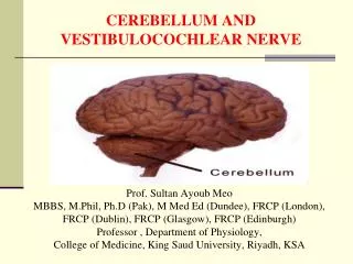
CEREBELLUM AND VESTIBULOCOCHLEAR NERVE Prof. Sultan Ayoub Meo MBBS, M.Phil, Ph.D (Pak), M Med Ed (Dundee), FRCP (London), FRCP (Dublin), FRCP (Glasgow), FRCP (Edinburgh) Professor , Department of Physiology, College of Medicine, King Saud University, Riyadh, KSA
Table 5.3 (1)Page 144 • Cerebral cortex • Cerebral cortex • Basal nuclei • (lateral to thalamus) • Basal nuclei • Thalamus • (medial) • Thalamus • Diencephalon • Hypothalamus • Hypothalamus • Cerebellum • Cerebellum • Midbrain(Mesencephalon) • Brain stem • (midbrain, pons, • and medulla) • Brain stem • Pons • Medullaoblongata • Spinal cord CEREBELLUM
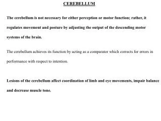
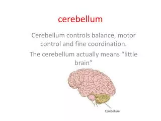
CEREBELLUM CEREBELLUM: Cerebellum is derived from a Latin word means "little brain.“ Cerebellum is the largest part of the hind brain, lies behind the pons and medulla Oblongata. Shape: Oval shaped, with an approximate weight is 150 gm Location: Situated in the posterior cranial fossa Anteriorly: 4th ventricle, pons, and medulla oblongata Superiorily: Covered by tentoriumcerebelli Posterio-inferiorly: Squamous occipital
CEREBELLUM The cerebellum is anatomically and physiologically divided into three parts: • Paleocerebellum: Anterior lobe [Spinocerebellum] • Neocerebellum: Posterior lobe [Cerebrocerebellum] • Archicerebellum: Flocculonodularlobe [Vestibulocerebellum]
CEREBELLAR PEDUNCLES: CARRY AFFERENTS FROM WHERE? Inputs to the Cerebellum from the cerebrum SuperiorCerebellar Peduncle Middle Cerebellar Peduncle Inputs to the Cerebellum from from the Pons Inferior Cerebellar Peduncle Inputs to the Cerebellum from the Medulla Oblongata
CerebellarPeduncles CEREBELLAR PEDUNCLES: CARRY AFFERENTS FROM WHERE? Three paired fiber tracts connect the cerebellum to the brainstem: • Superior peduncles connect the cerebellum to the cerebrum • Middle peduncles connect the cerebellum to the pons • Inferior peduncles connect the cerebellum to the medulla
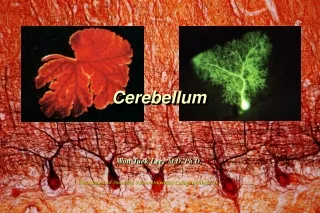
CEREBELLUM LAYERS The cerebellum has an external cerebellar cortex separated by white matter from the deep cerebellar nuclei as follows: Cerebellar Cortex Molecular Layer Purkinje Cell Layer Granular Layer Cerebellar Nuclei Dentate Nucleus Globose Nucleus Emboliform Nucleus Fastigial Nuclei Note: [Globose and Emboliform also known as interpositus nucleus Purkinje cells Basket cells Golgi cells GABA…Inhabi Granular cells Glutamate…Exci Stellate cells: Taurine…..Inhabi
NUCLEI OF THE CEREBELLUM DEEP NUCLEI 1. Fastigial nucleus 2. Globose nucleus 3. Emboliform nucleus 4. Dentate nucleus
OUTPUT FROM DEEP CEREBELLAR NUCLEI Fastigii Nucleus Interpositus Nucleus Dentate Nucleus Red Nucleus Premotor cortex Red Nucleus Motor Cortex Reticular Formation Control distal muscle during movement Planning of movement Its timing and initiation Control Axial muscle during movement
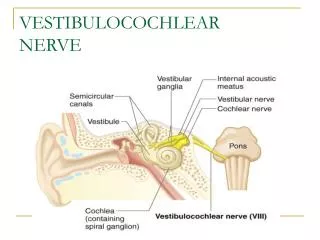
VESTIBULO COCHLEAR NERVE The vestibulo cochlear nerve conducts hearing (audition) and balance (vestibular). The receptor cells are located in the membranous labyrinth which is embedded in the petrous part of the temporal bone. There are two specialized organs in the bony labyrinth, the cochlea and the vestibular apparatus. The vestibular apparatus senses head position changes relative to gravity. Movement causes fluid vibration resulting in hair cell displacement that activates the vestibular part of the eighth nerve.
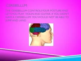
CEREBELLUM AND VOLUNTARY MOTOR CONTROL Cerebral and cerebellar control of voluntary movements, involving especially the intermediate zone of the cerebellum.
MAIN CONNECTIONS OF THE PALEOCEREBELLUM RED NUCLEUS NUCLEUS INTERPOSITUS Rubro spinal tract Inferior Olivry nucleus ANTERIOR LOBE PARAVERMAL ZONE Lower motor neuron PALEOCEREBELLUM SPINAL CORD Spinocerebellar tract
MAIN CONNECTIONS OF THE NEOCEREBELLUM CEREBRAL CORTEX THALAMUS DENTATE NUCLEUS pyramidal tract Pontine Nucleus POSTERIOR LOBE CEREBELLAR HEMISPHERE lower motor neuron NEOCEREBELLUM LMN
MAIN CONNECTIONS OF THE VESTBULOCEREBELLUM Vestibular Organ Floculonodular Lobe Vermis VESTIBULAR NUCLEUS vestibulospinal tract FASTIGIAL NUCLEUS MLF lower motor neuron ARCHICEREBELLUM LMN
CEREBELLUM AND AUTOMATIC MOTOR CONTROL CEREBELLUM Motor Cortex Red Nucleus Vestibular Nucleus Reticular Formation Lower Motor Neuron (LMN) Proprioceptors
Vestibulocerebellum • Spinocerebellum • Cerebrocerebelum CEREBELLUM Anterior Lobe Primary fissure It makes the movements smooth and coordinated Posterior Lobe It interacts with motor cortex in planning & programming of movements. Flocculo-Nodular Lobe (FN lobe) Maintenance of balance, control of eye movements • Folia
FINGER NOSE TEST While the examiner holds his finger at arm's length from the patient. Patient touches her nose and then touches the examiner's finger. After several sequences, the patient is asked to repeat the exercise with her closed eyes. A patient with a cerebellar disorder tends to miss the target.
DYSDIADOCHOKINESIS: RAPIDLY ALTERNATING MOVEMENTS Dysdiadochokinesis: Inability to perform rapidly alternating movements. Is called dysdiadochokinesia. It is usually caused by multiple sclerosis in adults and cerebellar tumors in children. Patients with other movement disorders (e.g. Parkinson's disease) may have abnormal rapid alternating movement testing secondary to akinesia or rigidity, thus creating a false impression of dysdiadochokinesia.
HEEL TO SHIN TEST The heel to shin test is a measure of coordination and may be abnormal if there is loss of motor strength, proprioception or a cerebellar lesion. If motor and sensory systems are intact, an abnormal, asymmetric heel to shin test is highly suggestive of an ipsilateralcerebellar lesion.
CEREBELLAR SIGNS Response delays Hypometria & Ataxia Incoordination/ rapid alternatingmovements (disdiadochokinesia)