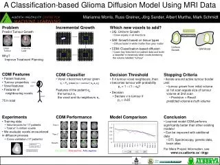

A Classification-based Glioma Diffusion Model Using MRI Data
E N D
Presentation Transcript
A Classification-based Glioma Diffusion Model Using MRI Data Marianne Morris1,2 Russ Greiner1,2, Jörg Sander2, Albert Murtha3, Mark Schmidt1,2 1Alberta Ingenuity Centre for Machine Learning 2 University of Alberta 3 Cross Cancer Institute, Alberta Cancer Board
Predict Tumour Growth Why? • Study tumour growth patterns • Improve treatment planning initial tumour tumour 6 months later
Outline • Introduction • Incremental Growth Modeling • Features • Models (UG, GW, CDM) • Experiments
Incremental Growth Model • Iteratively assign each voxel around the active tumour border totumour vsnon-tumour • Stops at termination condition • Reaching a specified size of tumour • … there’s no more voxels to add • Several Approaches
Incremental Growth Model Tumor
Incremental Growth Model Neighbours Tumor
Incremental Growth Model Tumor
Incremental Growth Model Neighbours Tumor
Incremental Growth Model Tumor
Incremental Growth Model Tumor
Which New Voxels to Add • UG: Uniform Growth • GW: Growth based on tissue types • CDM: Classification-based diffusion
Tumour growth modeling – uniform diffusion (UG) • Radial uniform growth (in all directions alike) Original tumour Final tumour volume
Tumour growth modeling – White vs. Grey matter (GW) • A 5:1 ratio for diffusion in white matter vs. grey matter (Sawnson et al., 2000) White matter Grey matter Original tumour Final tumour volume
Am I a tumour? Tumour growth modeling • Uniform growth: • Yes! • GW model: • If White matter: Yes! • If Grey matter: 20% • CDM model: • “Learn” tumour growth pattern voxel Active tumour border
Classification-Based Diffusion Model (CDM) • Preprocessing • Noise reduction • Spatial registration • Intensity Standardization • Tissue segmentation • Tumour segmentation • Feature extraction • Classification • Tumour growth modeling
tumour voxel patient Features • Patient features • Tumour properties • Voxel features • Features of neighbouring voxels A total of 75 features
Features: Patient • Age • Correlation between age and glioma grade(more aggressive tumours occur in older patients; benign tumours in children) patient
Features: Tumour • Area-volume ratio • Volume increase between 2 scans • Percentage of edema
Features: Voxel tumour voxel • Min Distance from tumour border • Tissue type derived from template • Tissue type derived from patient’s image • Image intensities (T1, T1-contrast, T2) • Template intensity • Edema region • Coordinates & Tissue Map • Distance-Area ratio edema tumour voxel tumour
Features: Neighbourhood y z 2 1 6 0 3 x 4 5 • For each of 6 neighbours* • Edema • Image intensities • Tissue type derived from template • Tissue type derived from patient’s image • A neighbourhood in 3D is the 6 voxels immediately adjacent to some voxel v (not including diagonal ones) 6 neighbours
Classification-Based Diffusion Model (CDM) • Preprocessing • Noise reduction • Spatial registration • Intensity Standardization • Tissue segmentation • Tumour segmentation • Feature extraction • Classification • Tumour growth modeling
tumour voxel v patient CDM Classifier • Voxel v becomes tumour given… qv = PΘ(class (v) = tumour | epatient,etumour,ev) Features of the patient epatient the tumour etumour the voxel and its neighbours ev
Learning Parameters (Classifier) • How to learn Θ ? • Naïve Bayes • Logistic Regression • Linear-kernel SVM • Trained on other brain images
Outline • Introduction • Incremental Growth Modeling • Experiments • Evaluation Measure • Model Comparison • Best Case • Average Case • Special Cases • Average P/R
Experimental Procedure • Training data • Sample of voxels in volume-difference between two scans including 2-voxel border around the volume at the 2nd time scan • Volume-pairs for 17 patients • Total of ½ million voxels • We evaluate voxels encountered in diffusion process • Cross-validation (17 patients) Original tumour Additional tumour growth
Tumour growth modeling – CDM (wrt Neighbours) • Voxel v becomes tumour based on… • Features: epatient,etumour,ev • Compute: qv = PΘ(class (v) = tumour | epatient,etumour,ev) • Neighbours of voxel v • If k tumour-voxel neighbours,probability that voxel vbecomes tumour pv = 1 – (1 – qv)k • Decision • Declare voxel v is tumour if pv0.65 v6,v7 : k = 0 v1,v2,v5 : k = 1 v3,v4 : k = 2
System Performance True positives False positives False negatives Left to right: Slices from lower to upper brain Time 1 scan Time 2 scan CDM prediction
Predicted pt Correct nt Evaluation • Precision, Recall • for tumour, non-tumour voxels nt = truth & pt = prediction ; Precision = Recall
Diffusion Modeling Process • We grow tumour from initial volume at 1st time scan to size of tumour volume at 2nd time scan • Precision = Recall because predicted volumetruth volume Tumour volume at 2nd time scan Tumour at 1st time scan
Results (Best case) GBM_7: CDM beats UG by 20% and GW by 12% Grew tumour along edema regions but… didn’t predict other wing of butterfly True positives False positives False negatives
Results (Average case) GBM_1: CDM beats UG by 6% and GW by 8% Need a more accurate brain atlas True positives False positives False negatives
Results (Special case) GBM_10: CDM beats UG by 8% and GW by 2% Resection & Recurrence True positives False positives False negatives
Results CDM performs significantly better than UG and GW! • T-test: the probability that the means are not significantly different • Paired data (same data sample; different models) • CDM vs UG: p = 0.001 • CDM vs GW: p = 0.001 • (UG vs GW: p = 0.034) X is the mean Var: the variance n: the number of samples
Future work • More expressive features • Spectroscopy, DTI, genetic data • Larger dataset (treatment effect) • Brain atlas (“highways” vs. “barriers”)
Conclusion • Challenge: Predicting how brain tumours will grow • Answer: Learned model CDM performs significantly better than other existing models! • … can improve with additional data
Acknowledgements • The University of Alberta;Dept of Computing Science • The Alberta Ingenuity Centre for Machine Learning • Cross Cancer InstituteAlberta Cancer Board • Brain Tumor Growth Prediction team