Download
1 / 54
660 likes | 1.66k Views
Lecture 7: Protein purification. Protein purification. Methods of solubilization. 1st step is to get the protein into solution If the protein is located in the cytosol, we need to break open the cell.

E N D
Lecture 7: Protein purification • Protein purification
Methods of solubilization • 1st step is to get the protein into solution • If the protein is located in the cytosol, we need to break open the cell. • For animal cells this can be accomplished with osmotic lysis. Put the cells in hypotonic solution; less solutes than inside the cell. • For cells with a cell wall (bacteria, plants) we have to use other methods. • For bacteria, lysozyme is often effective - Selectively degrades bacterial cell wall. • Can also use detergents or organic solvents, although these may also denature the protein.
Mechanical processes to break open the cell • High speed blender • Homogenzier • French press • Sonicator. After the cells have been broken, the crude lysate, may be filtered or centrifuged to remove the particulate cell debris. The protein of interest is in the supernatant.
For proteins that are components of membranes or subcellular assembly • Remove the assembly from the rest of the cellular material (mitochondria for example). • Can be done by differential centrifugation-cell lysate is centrifuged at a speed that removes only the cell components denser than the desired organelle followed by a centrifugation at speed that spins down the organelle.
Stabilization of proteins • pH • Temperature • Inhibition of proteases • Retardation of microbes that can destroy proteins • Sodium azide is often used.
Assay of proteins • Done throughout the purification process to make sure that your protein of interest is there. • If the protein of interest is an enzyme, using a reaction for which that enzyme is a catalyst is a good way to monitor protein recovery. • Monitor the increase of the product of the enzymatic reaction • Fluorescence • Generation of acid to be monitored by titration • Coupled enzymatic reaction - couple with another enzyme to make an observable substance.
Immunochemical techniques to assay for proteins • Use specific immunoglobulins (antibodies), proteins that interact specifically with the protein of interest and can be easily monitored. • Antibodies are produced by an animal’s immune system in response to the introduction of a foreign protein. • Antibodies specifically bind to the foreign protein. • Extracted from blood serum of animal that has been immunized against a particular protein. • Many different antibodies in sera with different specificities and binding affinities toward the protein of interest.
Immunochemical techniques to assay for proteins • Immune cells that produce antibodies normally die after a few cell divisions so it is difficult to get a specific antibody clone. • We can make monoclonal antibodies by fusing a cell producing the desired antibody with a cancer cell (myeloma). • This results in a hybridoma that is essentially an immortal cell, so large quantities of monoclonal antibody can be produced.
Figure 6-1 An enzyme-linked immunosorbent assay (ELISA). Page 130
Summary of initial steps of protein purification • Choose source of proteins. • Solubilize proteins. • Stabilize proteins. • Specific assay for protein of interest • Enzymatic activity, immunological activity, physical characteristics (e.g. molecular mass, spectroscopic properties, etc.), biological activity • Assay should be: • Specific • Rapid • Sensitive • Quantitative
Things to monitor during protein purification • Things to monitor during protein purification • Total sample volume • Total sample protein (est. by A280; 1.4-1.0 mg/ml) • Units of activity of desired protein (based on specific assay) • Other basic information to track • % yield for each purification step • Specific activity of the desired protein (units/mg of protein) • Purification enhancement of each step (e.g. “3.5 fold purification”) • In designing a purification scheme you have to balance purification with yield.
Solubilities of proteins • Multiple acid-base groups on proteins affect their solubility properties. • Solubility of a protein is therefore dependent on concentrations of dissolved salts, the polarity of solvent, the pH and the temperature. • Certain proteins will precipitate from solutions under conditions which others remain soluble-so we can use this as an initial purification step of proteins. • Salting out or salting in procedures take advantage of ionic strength 2 1/2ciZi Ionic strength (I) = Ci = molar concentration of ionic species Zi = ionic charge
Figure 6-2 Solubilities of several proteins in ammonium sulfate solutions. Page 131
Solubilities of proteins • A protein’s solubility at a given ionic strength varies with the types of ions in solution. • The order of effectiveness of these various ions in influencing protein solubility is quite similar for different proteins and is due to the ion’s size and hydration. • The solubility of a protein at low ionic strength generally increases with the salt concentration. This is called salting in. As the salt concentration increases the additional counterions more effectively shield the protein molecule’s multiple ionic charges and thereby increase the protein’s solubility. • At high ionic strengths the solubilities of proteins as well as most other substances decrease. This is called salting out and results from a competition between the added salt ions and the other dissolved solutes for molecules of solvation.
Figure 6-3 Solubility of caboxy-hemoglobin at its isoelectric point as a function of ionic strength and ion type. Page 131
Solubilities of proteins • Salting out is one of the most commonly used protein purification procedures. • By adjusting the salt concentration in a solution with a mixture of proteins to just below the precipitation point of the protein to be purified, many unwanted proteins can be eliminated from solution. Then after the precipitate is removed by filtration or centrifugation, the salt concentration of the remaining solution is increased to precipitate the desired protein. • Ammonium sulfate is the most commonly used reagent • High solubility (3.9 M in water at 0 ºC) • High ionic strength solution can be made (up to 23.5 in water at 0 ºC) Note-certain ions (I-, ClO4-, SCN-, Li+, Mg2+, Ca2+ and Ba+) increase the solubilities of proteins rather than salting out. (also denature proteins).
Solubilities of proteins • Water-miscible organic solvents also precipitate proteins. • Acetone, ethanol • Low dielectric constants lower the solvating power of their aqueouse solutions for dissolved ions. • This technique is done at low temperatures (0 ºC) because at higher temperatures, the solvent evaporates. • Can magnify the differences in salting out procedures. • Some water-miscible organic solvents (DMF, DMSO) are good at solubilizing proteins (high dielectric constants).
Solubilities of proteins • Proteins have various ionizable groups (many pK’s) • At a pH characteristic for each protein, the positive charges on the molecule exactly balance the negative charges (isoelectric point, pI). • At pI, the protein has no net charge and is immobile in an electric field. • Therefore, solubility can be influenced by changes in the pH.
Figure 6-4 Solubility of b-lactoglobin as a function of pH at several NaCl concentrations. Page 132
Solubilities of proteins • A protein in a pH near its isolectric point is not subject to salting in. • As the pH is moved away from the pI of the protein, the protein’s net charge increases and it is easier to salt in. • Salts inhibit interactions between neighboring molecules in the protein that promote aggregation and precipitation. • pI’s of proteins can be used to precipitate proteins.
Table 6-1 Isoelectric Points of Several Common Proteins. Page 133
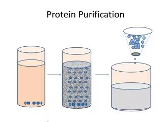
Column chromatography • After the initial fractionation steps we move to column chromatography. • The mixture of substances (proteins) to be fractionated is dissolved in a liquid or gaseous fluid called the mobile phase. • This solution is passed through a column consisting of a porous solid matrix called the stationary phase. These are sometimes called resins when used in liquid chromatography. • The stationary phase has certain physical and chemical characteristics that allow it to interact in various ways with different proteins. • Common types of chromatographic stationary phases • Ion exchange • Hydrophobic • Gel filtration • Affinity
Negatively charged (acidic) protein or enzyme - - - - Ion exchange chromatography Positively charged (basic) protein or enzyme • Ion exchange resins contain charged groups. • If these groups are acidic in nature they interact with positively charged proteins and are called cation exchangers. • If these groups are basic in nature, they interact with negatively charged molecules and are called anionexchangers. + CH2-COO- + CH2-COO- + + CM cellulose cation exchanger CH2-CH2 -NH+(CH2CH2) CH2-CH2 -NH+(CH2CH2) DEAE cellulose anion exchanger
CH2-COO- CH2-COO- CM cellulose cation exchanger Negatively charged proteins pass through the column - - - - - - - - Ion exchange chromatography For protein binding, the pH is fixed (usually near neutral) under low salt conditions. Example cation exchange column… Positively charged protein or enzyme bind to the column + + + +
CH2-COO- CH2-COO- CH2-COO- CH2-COO- + + CM cellulose cation exchanger CM cellulose cation exchanger + + + + + + Ion exchange chromatography To elute our protein of interest, add increasingly higher amount of salt (increase the ionic strength). Na+ will interact with the cation resin and Cl- will interact with our positively charged protein to elute off the column. + Increasing [NaCl] of the elution buffer Cl- Na+ Na+2 Cl- Na+ Cl- Cl- Na+2
Ion exchange chromatography • Proteins will bind to an ion exchanger with different affinities. • As the column is washed with buffer, those proteins relatively low affinities for the ion exchange resin will move through the column faster than the proteins that bind to the column. • The greater the binding affinity of a protein for the ion exchange column, the more it will be slowed in eluting off the column. • Proteins can be eluted by changing the elution buffer to one with a higher salt concentration and/or a different pH (stepwise elution or gradient elution). • Cation exchangers bind to proteins with positive charges. • Anion exchangers bind to proteins with negative charges.
Figure 6-6 Ion exchange chromatography using stepwise elution. Page 134
Ion exchange chromatography • Gradient elution can improve the washing of ion exchange columns. • The salt concentration and/or pH is continuously varied as the column is eluted so as to release sequentially the proteins bound to the column. • The most widely used gradient is the linear gradient where the concentration of eluant solution varies linearly with the volume of the solution passed. • The solute concentration, c, is expressed as c = c2 - (c2 - c1)f c1= the initial concentration of the solution in the mixing chamber c2= the concentration of the reservoir chamber f= the remaining fraction of the combined volumes of the solutions initially present in both reservoirs.
Figure 6-7 Device for generating a linear concentration gradient. Page 135 c = c2 - (c2 - c1)f
Figure 6-8 Molecular formulas of cellulose-based ion exchangers. Page 135
Ion exchange chromatography • Ion exchangers can be cellulosic ion exchangers and gel-type ion exchangers. • Cellulosic ion exchangers most common. • Gel-type ion exchangers can combine with gel filtration properties and have higher capacity. • Disadvantage-these materials are easily compressed so eluant flow is low. • There are other materials derived from silica or coated glass beads that address this problem.
Gel filtration chromatography • Also called size exclusion chromatography or molecular sieve chromatography. How does it work? If we assume proteins are spherical… Molecular mass (daltons) 10,000 30,000 100,000 size
Gel filtration chromatography • The molecular mass of the smallest molecule unable to penetrate the pores of the gel is at the exclusion limit. • The exclusion limit is a function of molecular shape, since elongated molecules are less likely to penetrate a gel pore than other shapes. • Behavior of the molecule on the gel can be quantitatively characterized. Total bed volume of the column Vt = Vx + V0 Vx = volume occupied by gel beads V0 = volume of solvent space surrounding gel; Typically 35%
Gel filtration chromatography • Elution volume (Ve) is the volume of a solvent required to elute a given solute from the column after it has first contacted the gel. • Relative elution volume (Ve/V0) is the behavior of a particular solute on a given gel that is independent of the size of the column. • This effectually means that molecules with molecular masses ranging below the exclusion limit of a gel will elute from a gel in the order of their molecular masses with the largest eluting first.
Figure 6-10 Molecular mass determination by gel filtration chromatography. Page 138
Table 6-3 Some Commonly Used Gel Filtration Materials. Page 138
Gel filtration chromatography • Elution volume (Ve) is the volume of a solvent required to elute a given solute from the column after it has first contacted the gel. • Relative elution volume (Ve/V0) is the behavior of a particular solute on a given gel that is independent of the size of the column. • This effectually means that molecules with molecular masses ranging below the exclusion limit of a gel will elute from a gel in the order of their molecular masses with the largest eluting first.
Affinity chromatography • Many proteins can bind specific molecules very tightly but noncovalently. • We can use this to our advantage with affinity chromatography. Glucose (small dark blue molecule) binding to hexokinase. The enzyme acts like a jaw and clamps down on the substrate (glucose)
Inert support Affinity chromatography • How does it work? • Ligand - a molecule that specifically binds to the protein of interest. Inert support + + Ligand Spacer arms Affinity material prepared