Download
1 / 31
320 likes | 571 Views
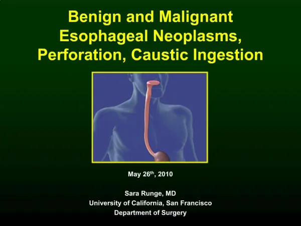
Benign and Malignant Esophageal Neoplasms, Perforation, Caustic Ingestion. May 26 th , 2010 Sara Runge, MD University of California, San Francisco Department of Surgery. Benign Neoplasms. Epithelial tumors papilloma, polyp, adenoma, cyst Nonepithelial tumors

E N D
Benign and MalignantEsophageal Neoplasms,Perforation, Caustic Ingestion May 26th, 2010 Sara Runge, MD University of California, San Francisco Department of Surgery
Benign Neoplasms • Epithelial tumors • papilloma, polyp, adenoma, cyst • Nonepithelial tumors • fibromyoma, leiomyoma, lipomyoma, fibroma, mesenchymal tumor, neurofibroma, osteochondroma • Heterotopic tumors • gastric mucosal tumor, melanoblastic tumor, sebaceous gland tumor, granular cell myoblastoma, pancreatic gland tumor, thyroid nodule
Leiomyoma • 2/3 of all benign tumors • Indications for resection: dysphagia, >5cm, increase in size, mucosa ulceration, rule out malignant process • Approach • <8cm: extramucosal enucleation • >8cm: esophageal resection
Malignant Neoplasms • Majority of esophageal cancer is squamous cell carcinoma or adenocarcinoma • Incidence of squamous cell cancer decreasing, while incidence of adenocarcinoma rising • Now equal incidence of each tumor in the U.S. • 17% overall 5 year survival rate
Risk Factors • Squamous cell carcinoma • Smoking • Alcohol • Foods containing N-nitroso compounds • Underlying esophageal disease (achalasia, caustic strictures) • Adenocarcinoma • Barrett’s esophagus • GERD • High BMI • Smoking
Malignant Transformation • Squamous cell carcinoma squamous epithelium -> epithelial dysplasia -> carcinoma in situ -> carcinoma • Adenocarcinoma squamous epithelium -> intestinal metaplasia to columnar epithelium (Barrett’s esophagus) -> low-grade dysplasia -> high-grade dysplasia -> adenocarcinoma
Clinical Manifestations • Dysphagia to solids (80%) • Weight loss (50%) • Odynophagia (20%)
Staging • T T1: Invades lamina propria, muscularis mucosa, or submucosa T2: invades muscularis propria T3: invades adventitia T4: invades adjacent structures • N N1: 1-2 regional LNs N2: 3-6 regional LNs N3: 7+ regional LNs • M M1: distant mets
Diagnosis • Endoscopy and biopsy • CT chest/abdomen • PET- to detect mets • Endoscopic ultrasound- most accurate for locoregional tumor staging
Treatment • Only 30-40% resectable at presentation • Cervical esophageal cancer usually treated like other SCC of head/neck… chemorad preferred over surgery • Thoracic esophageal cancer requires total esophagectomy due to risk of submucosal skip lesions
Superficial esophageal cancer • Esophagectomy • Endoscopic resection • For Tis (high-grade dysplasia) or T1a (invades mucosa but not submucosa) • If specimen shows deeper invasion, can then get esophagectomy • RFA, photodynamic therapy • For dysplastic Barrett’s epithelium • Disadvantage: no specimen
Transhiatal esophagectomy • Exposure with upper midline laparotomy and left neck incision • Thoracic esophagus bluntly dissected through each incision • Cervical anastomosis created utilizing gastric pull-up • Disadvantages: inability to perform full thoracic lymphadenectomy, lack of visualization of mid-thoracic dissection
Ivor-Lewis (transthoracic) esophagectomy • Exposure with laparotomy and right thoracotomy • Intrathoracic anastomosis • Advantages: direct visualization of thoracic esophagus, can perform full lymphadenectomy, lower rate of anastomotic leak • Disadvantages: greater likelihood of bile reflux
Three-hole esophagectomy • Exposure with right posterolateral thoracotomy, laparotomy, left neck incision • Advantages: direct visualization of thoracic esophagus, can perform full lymphadenectomy, lower rate of bile reflux
Adjuvant Therapy • Timing (pre-operativey vs post-operatively) and type (chemo vs rad vs chemorad) controversial • Surgery alone perferred for stage I • Neoadjuvant chemorad followed by surgery preferred for stages IIB & III if distal esophageal or GE junction cancer • Patients with completely resected node-positive cancer who haven’t received neoadjuvant therapy should get adjuvant chemo or chemorad
Locally advanced unresectable cancer • Potentially resectable (T4a)- invasion of pleura, pericardium, diaphragm • Unresectable (T4b)- invasion of aorta, trachea, heart, great vessels, presence of tracheoesophageal fistula • Neoadj chemorad may make unresectable disease resectable
Locally advanced unresectable cancer • Radiation +/- chemo • Esophageal dilatation +/- stenting • Photodynamic therapy, laser ablation
Caustic Ingestion • Ingestion of strong acids or strong bases • First step is airway protection... may need tracheostomy • Endoscopy • Minor/questionable ingestion: endoscopy after 24-48 hrs • Large ingestion: immediate endoscopy to guide treatment
Caustic Ingestion SEQUELAE: • Esophageal stricture • 1/3 of patients with caustic injury develop strictures (more likely if higher grade injury) • treat with repeat dilations • Esophageal squamous carcinoma • 1000x higher risk compared to general population • usually alkali ingestion • begin surveillance 15-20 yrs after exposure wtih endoscopy every 1-3 yrs
Esophageal perforation CAUSES: • Instrumental (59%) • Endoscopy • Dilation • Intubation • Noninstrumental • Swallowed foreign body (12%) • Penetrating neck/chest/abd trauma (9%) • Corrosive injuries • Boerhaave's syndrome
Esophageal perforation CLINICAL FEATURES: • Cervical: cervical dysphagia, neck pain, dysphonia, subcutaneous cervical emphysema • Intrathoracic: symptoms of mediastinitis (tachycardia, tachypnea, fever, leukocytosis) • Intraabdominal: symptoms of acute abdomen (tachycardia, tachypnea, fever, leukocytosis)
Esophageal perforation DIAGNOSIS: • Contrast esophagram with gastrografin (gold standard) • repeat with barium if negative with gastrografin • Flexible endoscopy • to help localize for OR planning • contraindicated if suspect mucosal tear -> can cause full-thickness tear • CT- to localize fluid collections
Esophageal perforation MANAGEMENT:
Esophageal perforation MANAGEMENT: • Upper third: cervical drainage • left neck incision, G or J tube for enteral feeding • Middle third: • right 5th intercostal thoracotomy, G or J tube, chest tubes • buttressed primary repair w/ flaps of pleura, pericardium, diaphragm, omentum, muscle • Lower third: • left 7th intercostal thoracotomy, G or J tube, chest tubes • buttressed primary repair w/ pedicled intercostal muscle flap
References • Brinster CJ, et. al. Evaluation and treatment of esophageal perforation. Ann Thorac Surg. 2004; 77:1475. • Cameron, JL. Current Surgical Therapy. 8th Edition. Philadelphia: Mosby; 2004. • Mulholland MW. Greenfield's Surgery. 4th Edition. Philadelphia: Lippincott Williams & Wilkins; 2006. • Paidas, CN. Caustic burns of the esophagus. Current therapy in thoracic and cardiovascular surgery. Philadelphia: Mosby; 2004:99.