Download
1 / 47
480 likes | 631 Views
C SPINE. Y A Mamoojee. Importance of Prompt Diagnosis. Neck pain > quadriplegia > death Delayed recognition can lead to irreversible s.c injury and permanent neurologic damage. INDICATIONS. Who needs XR. NEXUS. NO - Alcohol intoxication Focal neuro deficit Midline tenderness

E N D
C SPINE Y A Mamoojee
Importance of Prompt Diagnosis • Neck pain • > quadriplegia • > death • Delayed recognition can lead to irreversible s.c injury and permanent neurologic damage.
INDICATIONS • Who needs XR
NEXUS NO - • Alcohol intoxication • Focal neuro deficit • Midline tenderness • GCS 15 • Painful distracting injuries
CASE DISCUSSION • A person arrives by ambulance to ED on a backboard and a cervical collar after an MVA. • Speed of 50km/hr • No LOC, no other injuries, no midline tenderness, BAL 0.20. • Does he need imaging?
LATERAL • AP • ODONTOID • SWIMMERS • FLEXION/EXTENSION?
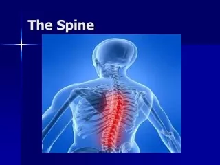
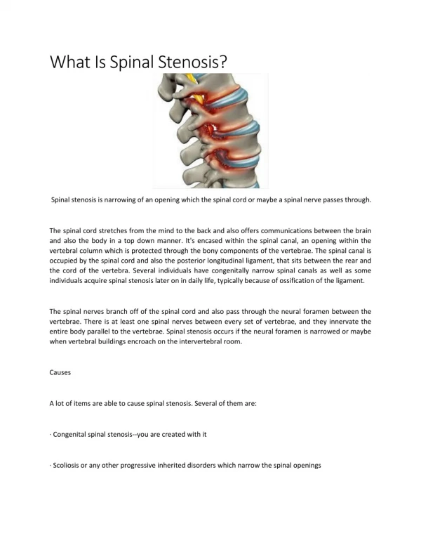
ANATOMY OF NECK • LIGAMENTS • BONES • MUSCLES • JOINTS
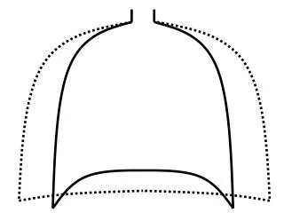
Most important view • Can see 80-90% of injuries • Interpretation: • A - adequacy • A - alignment • B - bone • C - cartilage • D - disc • S – soft tissue • A - Must have a view of C7 – T1 • A - Use 3 lines • 1. anterior vertebral line • 2. posterior vertebral line • 3. spino laminar line (base of spinous processes) • 4th line can be used ie. Tips of spinous processes
Check : • B - individual vertebrae • C - cartilage • D - disc • S - soft tissue - • <7mm at C3 • <21mm at C7 • no more than vertebral body width at C7 • Predental space – • 5mm child • 3mm adult • Fanning of spinous processes
Open mouth view • Adequate if entire Odontoid and lateral borders of C1 and C2 visible • Check : • lateral masses of C1 must align with Odontoid • bilateral symmetry • Important also for Odontoid fractures
SWIMMER’S AP
MECHANISM OF INJURY • 1. Flexion • 2. flexion rotation • 3. extension • 4. axial compression • 5. Other
WEDGE FRACTURE • STABLE • Compression fracture resulting from flexion • Features – • Buckled anterior cortex • Loss of height of anterior part of body • Anterosuperior fracture of vertebral body
FLEXION TEARDROP FRACTURE • UNSTABLE • Posterior ligament disruption and anterior compression fracture of the vertebral body • Prevertebral swelling • Tear drop fragment • Posterior vertebral body subluxation into the spinal canal • Spinal cord compression • Fracture of spinous process
Mechanism – Hyperflexion and Compression – Excessive flexion of the neck in the sagittal plane, disrupts posterior ligament. • Example – diving into shallow pool
ANTERIOR SUBLUXATION • Disruption of the posterior ligament complex. Anterior subluxation of C4 on C5 is characterized by widening of the interspinous space (arrowhead), subluxation of the C4-C5 interfacetal joints (arrows), and anterior rotation of the C4 vertebra relative to C5.
Stable but potentially unstable during flexion • Mechanism : hyperflexion • Disruption of posterior ligament complex, anterior intact • Stable – • loss of normal cervical lordosis • anterior displacement of body • fanning of interspinous distance • Unstable – • anterior subluxation >4mm • assoc. compression fracture >25% of affected body • increase or decrease in normal disc space • fanning of interspinous distance
BILATERAL FACET JOINT DISLOCATION • Complete anterior dislocation of the vertebral body • Mechanism – extreme hyperflexion of head and neck without axial compression • Unstable – very high risk of cord damage • Features – • complete anterior dislocation >50% of vertebral body diameter • Disruption of the posterior ligament complex and anterior longitudinal ligament • “Bow tie” appearance of the locked facets.
CLAY SHOVELLER’S FRACTURE • Fracture of spinous process C6-T1 • Mechanism – powerful hyperflexion, usually combined with contraction of paraspinous muscles pulling on spinous processes (e.g. shovelling). Features – spinous process fracture on lateral view Ghost sign on AP – double spinous process of C6/C7 due to displaced fractured spinous process
UNILATERAL FACET JOINT DISLOCATION • Stable • Mechanism – simultaneous flexion and rotation • Facet joint dislocation and rupture of the apophyseal joint ligaments • FEATURES : • Anterior dislocation of vertebral body by <50% of the diameter • Discordant rotation above and below involved level • Facet within intervertebral foramen on oblique view • “Bow tie” appearance of the overriding locked facets
EXTENSION INJURIES • Excessive extension of the neck in the sagittal plane. • E.g. hitting the dash board in MVA
HANGMAN’S FRACTURE • Fractures through pars interaticularis of the axis • Unstable if occurs with facet dislocation • Mechanism – hyperextension • Features – • Prevertebral soft tissue swelling • Avulsion of anterior inferior corner of C2 assoc. with rupture of the ant. Longitudinal ligament • Anterior dislocation of C2 body • Bilateral C2 pedicle fractures.
C1 POSTERIOR ARCH FRACTURE • Hyperextended head • C1 arch is compressed by occiput and C2 spinous process • Odontoid process is normal • Stable • Distinguish from Jefferson fracture (unstable)
BURST FRACTURE • Fracture of C3-C7 that results from axial compression • Spinal cord injury secondary to displacement of posterior fragments is common. • Mechanism – Axial compression • >25% loss of height of vertebral body • Stable • Needs CT or MRI
JEFFERSON FRACTURE • Burst type fracture of C1 • Lateral displacement of C1 masses • Fracture of anterior and posterior arches on both sides – quadruple fracture • Unstable – transverse ligament rupture • Soft tissue swelling is marked on Xray
ATLANTO AXIAL SUBLUXATION • Flexion and rotation causes the transverse ligament to rupture • Predental space >3.5mm in adults and >5mm in children • Unstable
ODONTOID FRACTURES • 3 Types : • I Avulsion of tip at alar ligament (stable) • II Base of dens (unstable) – common, non union is a complication • III Involves body of C2 (unstable)