Download
1 / 41
410 likes | 635 Views
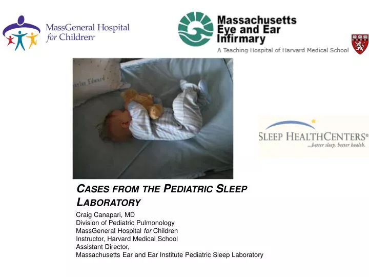
Cases from the Pediatric Sleep Laboratory. Craig Canapari, MD Division of Pediatric Pulmonology MassGeneral Hospital for Children Instructor, Harvard Medical School Assistant Director, Massachusetts Ear and Ear Institute Pediatric Sleep Laboratory. Overview.

E N D
Cases from the Pediatric Sleep Laboratory Craig Canapari, MD Division of Pediatric Pulmonology MassGeneral Hospital for Children Instructor, Harvard Medical School Assistant Director, Massachusetts Ear and Ear Institute Pediatric Sleep Laboratory
Overview • Why children are different; how these differences affect gathering and interpreting data in the lab • Consideration of the respiratory system • Cases • Summary
Stuff to know about kids • They are small and ornery • You have to deal with the parents too
Patients with “unusual” medical conditions are common • Seizure disorders and abnormal EEG • Underlying lung disease: prematurity, chronic aspiration, interstitial lung disease • Defects of the respiratory pump: neuromuscular and chest wall disorders • Disorders of respiratory control: central congenital hypoventilation syndrome • Patients with various syndromes • Additional monitoring is mandatory: video, end tidal CO2, extra EEG leads if indicated • Normative values are different; less well validated; unclear
AIRWAY: OSA CO2 Oxygenation RM Schwartzstein, Respiratory Physiology: A Clinical Approach, 2006
Capnography • B: Dead space ventilation • B - C: Ascending expiratory phase • C - D: Alveolar Plateau • D: End-tidal CO2D - E: Descending inspiratory phase
Disorders of Breathing Control: Central Congenital Hypoventilation Syndrome Characterized by a lack of respiratory effort. Usually neurological in origin.
Disorders of Respiratory Control • Ondine’s curse • Brainstem malformation e.g. Arnold-Chiari malformation • Central congenital hypoventilation syndrome (CCHS)
CCHS • Associated with lack of response to hypoxemia or hypercarbia especially during NREM sleep • Gene identified (repeats in PHOX2b) • Usually presents in infancy • Therapy • Invasive vs. NIPPV • Diaphragmatic pacing • Passive leg movements
CCHS Hypoventilation during NREM sleep Lack of heart rate variability Treated on PPV PC 19/5 x 15
Down syndrome • Flat facies or midfacial hypoplasia • Enlarged tongue (glossoptosis) • Low tone • Mental retardation • Risk of aspiration
Craniofacial considerations Micrognathia Cummings, Otolaryngology: Head & Neck Surgery, 4th ed. Midfacial hypoplasia http://www.chw.org/display/PPF/DocID/35350/router.asp
Normal Midfacial hypoplasia Ferraro, N. “Craniofacial development and the airway during sleep” in Sleep and Breathing in Children, 2nd ed. 2008
Polysomnographic patterns in Down syndrome • Hypoxemia • Hypoventilation • Central apneas • Increased risk of seizures • Severe OSA
Decreased reserve in a child with Trisomy 21 History of chronic aspiration; borderline SpO2 becomes abnormal at transition to REM.
Prader-Willi Syndrome Characteristic hypotonia in infancy and severe obesity in childhood
Prader-Willi Syndrome • Low tone and failure to thrive in infancy • Uncontrollable appetite from early childhood on results in severe obesity • Hypothalamic dysfunction • Sleep issues • Excessive daytime sleepiness common • Decreased peripheral chemoreception • Decreased response to hypoxemia, pH, elevated CO2 • Central and obstructive sleep apnea • (70% prevalence of OSA)
Prader-Willi Syndrome and Growth Hormone Treatment • Children with PWS have increased tone and energy with GH treatment • Rare cases of sudden death associated with growth hormone therapy • PSG is recommended prior to therapy and within one month of starting • Risk markers unclear, however
Central apneas? Sensitivity of belts at 50
Sensitivity of abdominal belt increased to 10: obstructive event
Duchenne Muscular Dystrophy • X-linked mutation of dystrophin gene • Affected boys present with delayed motor milestones • Wheelchair bound in second decade • Respiratory muscle weakness • Recurrent pneumonia • Hypoventilation
Sleep manifestations • OSA • Frequent repositioning • Hypoventilation • Silent • Nocturnal/AM headaches and nausea • Disrupted sleep • Breathlessness • Annual polysomnography is recommended once using a wheelchair full time
Minute ventilation = Tidal volume x respiratory rate Decreased ventilation = increased CO2 Tidal volume may be reduced by decreased muscle strength, chest wall deformity, obesity, upper airway obstruction RM Schwartzstein, Respiratory Physiology: A Clinical Approach, 2006
BiPAP/NIPPV • Bilevel Positive Pressure ventilation is the most common type of non-invasive positive pressure ventilation • In obstruction: • EPAP to get rid of apneas • IPAP to get rid of hypopneas • Keep span 4-6 • In hypoventilation • Keep EPAP low • Titrate IPAP to normalize gas exchange in REM • Pediatric patients may require a back up rate
Bilevel: gas exchange normalized Persistent asynchrony; back up rate added
Fibrodysplasia progressiva ossificans: progressive spinal deformity and restrictive lung disease.
Why not just use oxygen? • In patients with chronic ventilatory failure, supplemental oxygen may be associated with a severe rise in PaCO2. • Why? • Dogma: • Patients will STOP BREATHING if you give them oxygen because they are dependent on their hypoxic ventilatory drive to breathe • The story is a bit more complicated • CO2 corrosive to sleep quality *In our practice, CF and NM disorders. On the adult side, COPD
Causes of increased CO2 with O2 therapy. • Increase in dead space ventilation secondary to loss of hypoxic vasonconstriction (in context of PaO2< 60 torr-- O2 sat ~90% or less.) [48%] • Haldane effect. [30%] • Decrease in minute ventilation. [22%]
Oxygen in a child with DMD 0.5 L/min NC From MH Wagner, Journal of Clinical Sleep Medicine 4(2) 2008.
Caveats • If a patient needs O2, give it but know how much you are giving • Venturi mask • Target sats 88%-93% • If concerned, follow ABGs. • NIPPV may be preferable
Respiratory Control: Down Syndrome DMD PWS CCHS RESPIRATORY CONTROL ISSUES: Down Syndrome DMD PWS CCHS AIRWAY: Down Syndrome DMD VENTILATORY PUMP ISSUES: DMD Chest wall disease Obesity? CO2 GAS EXCHANGE ISSUES: Chronic Aspiration Chronic Lung disease of prematurity Interstitial lung disease RM Schwartzstein, Respiratory Physiology: A Clinical Approach, 2006
Summary • Pediatric syndromes may present with impairment in single or multiple domains of breathing • Insights obtained from these patients gives insight into more common pathophysiologic processes such as OSA, obesity, etc.
Thanks! Ellen Grealish Sheila Rubino Effie Chrysovergis Anita Rondinelli Adriana Rondinelli Rebecca Doe Melissa Kiernan