Download
1 / 18
290 likes | 894 Views
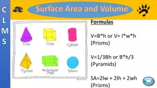
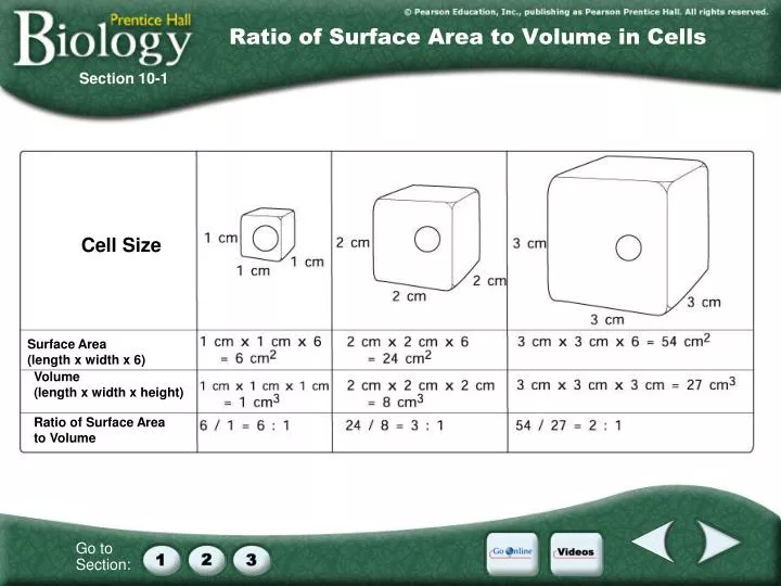
Ratio of Surface Area to Volume in Cells. Section 10-1. Cell Size. Surface Area (length x width x 6). Volume (length x width x height). Ratio of Surface Area to Volume. Figure 10–4 The Cell Cycle. Section 10-2. G 1 phase. M phase. S phase. G 2 phase.

E N D
Ratio of Surface Area to Volume in Cells Section 10-1 Cell Size Surface Area (length x width x 6) Volume (length x width x height) Ratio of Surface Area to Volume
Figure 10–4 The Cell Cycle Section 10-2 G1 phase M phase S phase G2 phase
In between divisionsCells are in this phase most of the time Can see nucleus DNA spread out as chromatin Can’t see chromosomes DNA gets copied (S) Cell gets ready to divide INTERPHASE (G1 - S - G2)
Pearson Education Inc publishing as Pearson Prentice Hall PROPHASE 1st dividing phase http://www.life.uiuc.edu/plantbio/102/lectures/08mit&veg102.html DNA scrunches into chromosomes Centrioles appear in centrosome region & move to poles Nuclear membrane & nucleolus disappear Spindle fibers form & attach to chromosomes
CENTROSOME ________ region organizes spindle Spindle MICROTUBULES are part of cytoskeleton http://www.coleharbourhigh.ednet.ns.ca/library/organelle_worksheet.htm
Chromosomes line up in ___________ middle METAPHASE Images from:Pearson Eduction Ince; Publishing as Pearson Prentice Hall http://www.science.siu.edu/plant-biology/PLB117/JPEGs%20CD/0247.JPG
Centromeres splitCentrioles pull chromatids_______ apart ANAPHASE Images from:Pearson Eduction Ince; Publishing as Pearson Prentice Hall http://www.science.siu.edu/plant-biology/PLB117/JPEGs%20CD/0247.JPG
two See ______ nuclei Nuclear membrane & nucleolus return TELOPHASE (reverse prophase steps) Chromosomes spread out as chromatin Centrioles disappear Spindle fibers disappear Images from:Pearson Eduction Ince; Publishing as Pearson Prentice Hall http://www2.bc.cc.ca.us/cnewton/Biology%2011/Mitosis.html
CYTOKINESIS Cytoplasm splits into 2 cells ANIMAL CELLS pinch cytoplasm in two with a ______________________ CLEAVAGE FURROW
CYTOKINESIS Cytoplasm splits into 2 cells PLANT CELLS can’t pinch because they have a sturdy ____________ Plant cells separate cytoplasm by growing a _______________ down the middle. CELL WALL CELL PLATE http://www.eastcentral.edu/acad/depts/BI/plant_mitosis_nolabels.html
Figure 10–5 Mitosis and Cytokinesis Section 10-2 Spindle forming Centrioles Centromere Chromatin Centriole Nuclear envelope Chromosomes (paired chromatids) Interphase Prophase Spindle Cytokinesis Centriole Metaphase Individual chromosomes Telophase Anaphase Nuclear envelope reforming
Figure 10–5 Mitosis and Cytokinesis Section 10-2 Spindle forming Centrioles Centromere Chromatin Centriole Nuclear envelope Chromosomes (paired chromatids) Interphase Prophase Spindle Cytokinesis Centriole Metaphase Individual chromosomes Telophase Anaphase Nuclear envelope reforming
Figure 10–5 Mitosis and Cytokinesis Section 10-2 Spindle forming Centrioles Centromere Chromatin Centriole Nuclear envelope Chromosomes (paired chromatids) Interphase Prophase Spindle Cytokinesis Centriole Metaphase Individual chromosomes Telophase Anaphase Nuclear envelope reforming
Figure 10–5 Mitosis and Cytokinesis Section 10-2 Spindle forming Centrioles Centromere Chromatin Centriole Nuclear envelope Chromosomes (paired chromatids) Interphase Prophase Spindle Cytokinesis Centriole Metaphase Individual chromosomes Telophase Anaphase Nuclear envelope reforming
Figure 10–5 Mitosis and Cytokinesis Section 10-2 Spindle forming Centrioles Centromere Chromatin Centriole Nuclear envelope Chromosomes (paired chromatids) Interphase Prophase Spindle Cytokinesis Centriole Metaphase Individual chromosomes Telophase Anaphase Nuclear envelope reforming
Figure 10–5 Mitosis and Cytokinesis Section 10-2 Spindle forming Centrioles Centromere Chromatin Centriole Nuclear envelope Chromosomes (paired chromatids) Interphase Prophase Spindle Cytokinesis Centriole Metaphase Individual chromosomes Telophase Anaphase Nuclear envelope reforming
Concept Map Section 10-2 Cell Cycle includes is divided into is divided into
M phase (Mitosis) Interphase G1 phase S phase G2 phase Prophase Metaphase Anaphase Telophase Concept Map Section 10-2 Cell Cycle includes is divided into is divided into