Download

1 / 65
700 likes | 1.23k Views
The Endocrine System. Specialist: Endocrinologist. Hormones. A single hormone can initiate many different cellular responses in different cells Hormones released in one part of the body , travel through the blood and regulate activity of cells in another part of the body

E N D
The Endocrine System Specialist: Endocrinologist
Hormones • A single hormone can initiate many different cellular responses in different cells • Hormones released in one part of the body, travel through the blood and regulate activity of cells in another part of the body • Some act within seconds, most within several minutes, some take hours or days to onset of action. • Duration of action can be seconds to days
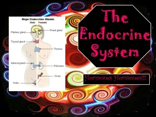
Strictly Endocrine Glands • Pineal • Melatonin • Pituitary • hGH, FSH, LH, TSH, • Thyroid • T3, T4 • Parathyroid • PTH • Adrenal • Aldosterone • Cortisol • Androgens • Epinephrine • Norepinephrine
Other Tissues/ organs that secrete hormones • Some tissues or organs are not strictly endocrine glands and have other functions, in addition to secreting hormones. • Hypothalamus • Thymus • Pancreas • Ovaries • Testes • Kidneys • Stomach • Liver • Small intestine • Skin • Heart • Adipose • placenta
Endocrine Glands are highly vascularized • Endocrine glands are very vascularized • Gland cells secrete their products into the interstitial fluid • The hormone then diffuses into the blood which carries it all over body • Only cells with receptors for that hormone will react with an action
Hormone Receptors • Lipid soluble hormones, like steroids & thyroid hormone, affect cell function by entering the nucleus and directly altering gene expression • Water soluble hormones activate plasma membrane receptors, which elicit second messengers that activate various enzymes inside the cells
Chemical Classes of Hormones Lipid Soluble Hormones: bind to receptors inside target cells Water Soluble Hormones: bind to receptors on target cell surface Amines Catecholamines: epinephrine, norepinephrine, dopamine - adrenal medulla Melatonin - pineal gland Histamine – mast cells in CT Serotonin – platelets Peptides & Proteins Many, including: all hypothalamic releasing and inhibiting hormones; all pituitary hormones, such as oxytocin, ADH, FSH, LH, TSH, ACTH, HGH, MSH; insulin, glucagon, somatostatin, Eicosanoids Eg Prostaglandins, Leukotrienes • Steroids • Aldosterone, cortisol, androgens – Adrenal cortex • Calcitriol- Kidneys • Testosterone - Testes • Estrogen, Progesterone – Ovaries • Thyroid • T3 (triiodothyronine) • T4 (thyroxine) • Gases • Nitric Oxide
Peptides, Amines, Steroids • Water soluble peptides or amines bind to receptors on the surface of cells • Lipid soluble hormones diffuse into the cell and bind receptors inside the cell
FYI - 2ndmessengers: cAMP • Water soluble hormones bind to surface receptors, & then activate a G protein. • The G protein, then activates an enzyme, AdenylateCyclase. • AdenylateCyclasecatalyzes conversion of ATP into the 2nd messenger, cAMP (cyclic AMP) • cAMP activates the enzyme, protein kinase, which can phosphorylate (add a phosphate) various proteins to activate them & produce a cellular action eg glycogen breakdown
FYI - 2ndmessengers: DAG & iP3 • Another example of a 2nd messenger. • Activated G protein can then activate a different enzyme, Phospholipase C (PLC) • PLC can activate both 2nd messengers, DAG & iP3… • Binding of a single epinephrine molecule can activate 100 G proteins • Each adenylcyclate can produce 1000 cAMP…
Control of Hormone secretion Hormone secrection is controlled by • signals from the nervous system • chemical changes in blood • Other hormones • Negative feedback
Hypothalamus The main link between the nervous and endocrine system
Hypothalamic Neurosecretory cells • Neurons in the hypothalamus are called Neurosecretorycells. These neurons of the hypothalamus secrete: • Releasing Hormones, such as PRH (Prolactin Releasing Hormone), or TRH (Thyroid Releasing Hormone). • Inhibiting Hormones, such as PIH (Prolactin Inhibiting Hormone).
Releasing and Inhibiting Hormones enter the Pituitary • The hypothalamic neurons secrete their releasing and inhibiting hormones into a capillary network on the infundibulum, or pituitary stalk. • The hormones then enter a second capillary network on the anterior pituitary via the hypophyseal portal vein. • Longer hypothalamic neurons reach the separate capillary network of the posterior pituitary directly. • Releasing hormones exert their effects on the cells of the pituitary and cause them to release pituitary hormones into the blood.
Anterior Pituitary Hormones Anterior pituitary endocrine cells produce the following anterior pituitary hormones: • Somatotrophs produce Human Growth Hormone (hGH) • Lactotrophs produce Prolactin (PRL) • Corticotrophs secrete Adrenocorticotropic Hormone (ACTH) & Melanocyte Stimulating Hormone (MSH) • Thyrotrophs secrete Thyroid-Stimulating Hormone (TSH) • Gonadotrophs synthesize Follicle Stimulating Hormone (FSH) & Luteinizing Hormone (LH)
Posterior Pituitary: 1) ADH& 2) Oxytocin • Hormones of the posterior pituitary are released directly from neurons that originate in the hypothalamus. • The neurosecretory cells make Anti Diuretic Hormone (ADH) and Oxytocin. They release them into the blood from their axon terminals when stimulated to do so.
Feedback Control of Pituitary Hormones • The pituitary and hypothalamus have receptors for each of the hormones they stimulate production of. • When blood levels of these hormones rise above a certain amount, the hypothalamus and pituitary will stop producing releasing hormones or pituitary hormones. • This is called negative feedback.
INSULIN LIKE GROWTH FACTOR (IGF-1) GROWTH HORMONE
Human Growth Hormone & Insulin Like Growth Factor • hGH is most plentiful anterior pituitary hormone. • When the pituitary secretes hGH, it stimulates the liver and other tissues to synthesize and secrete the protein hormone, IGF, insulin-like growth factor. • IGF is also called somatomedin.
IGF METABOLISM • Stimulates growth by increasing uptake of amino acids and speeding up protein synthesis. // Decreases breakdown of proteins into amino acids for ATP production. • Enhances lipolysis, using FA for ATP synthesis. • Raises blood sugar by stimulating liver to make glucose EFFECTS • In children, promotes growth of skeleton and skeletal muscles • In adults maintains muscle and bone mass, promotes tissue healing and repair
HYPOglycemiastimulates GHRH • Low glucose (hypoglycemia) stimulates the hypothalamus to release GHRH • GHRH stimulates the pituitary to secrete hGH • hGH stimulates the liver and other tissues to release of IGF
HYPERglycemiastimulates GHRIH • High IGF-1 or high blood glucose, stimulates the hypothalamus to secrete Growth Hormone Release InhibitingHormone (GHRIH), also known as somatostatin (SRIF) • GHRIH inhibits pituitary secretion of hGH • Low IGF slows glycogen breakdown in the liver & glucose enters the bloodstream more slowly
TRH, THS, T3, T4 thyroid
Thyroid Follicles • The thyroid is located below the cricoid cartilage of the larynx • It is made up of spherical sacs called thyroid follicles • Follicular cells surround a lumen filled with sticky fluid colloid, made of thyroglobulin and iodine • Parafollicular cells or C cells produce calcitonin
Synthesis of T4 & T3, Iodide & TGB • Follicular cells do the following: • Actively trap iodide ions from the blood into the cytosol • Synthesize Thyroglobulin (TGB). Adds tyrosine rings, and releases it into the lumen of the follicle • The enzyme, Thyroid Peroxidase, TPO oxidizes iodide oxidized (removes an electron) to make it iodine and then couples it to tyrosine on thyroglobulin to create MIT, Monoiodotyrosine, DIT, Diiodotyrosine, T3 & T4.
Coupling & Secretion of T3 & T4 • MIT or DIT will couple, joining to becomeT3 or T4 • Droplets of colloid re-enter the follicular cell by pinocytosis. • Lysosomes break down thyroglobulin, cleaving off T3 & T4 • Lipid soluble T3 & T4 diffuse out of the follicular cell into blood
Transport protein: TBG • >99% of T3&T4 combines with TBG, ThyroxineBinding Globulin, a transport protein that carries thyroid hormone in the blood
T4 to T4 conversion • Most body cells have receptors for thyroid hormone • T4 is converted to the more potent, T3 in the body tissues by deiodinase enzymes • 60% of this conversion occurs in the liver
Thyroid hormone targets & effects EFFECTS OF T3: • Increases Basal Metabolic Rate ie rate of ATP synthesis by mitochondria • Uses glucose and fatty acids for metabolism • Increases lipolysis and reduces cholesterol • Increases protein synthesis • Increases synthesis of Na/K/ATPase pumps • Upregulates beta receptors, thus increases effects of Epi & NE
Thyroid Feedback inhibition • The hypothalamus and pituitary glands will detect elevated amounts of T3 and T4 • They will reduce production of TRH & TSH
Parafollicular cells calcitonin
C cells/ parafollicular cells • Elevated blood Ca2+ stimulates cells in between the follicles, called C cells or Parafollicularcells • These cells secrete the hormone, calcitonin • Calcitonin decreases Ca and Phosphates in the blood by inhibiting osteoclasts • Accelerates uptake of Ca & Phosphate by bone
PTH Increases blood Ca2+ & Mg2+ parathyroid
Parathyroid • Decreased Ca2+ levels stimulate parathyroid chief cells to secrete PTH, parathyroid hormone • This gland is a major regulator of Ca, Mg, HPO42- (phosphate)
Parathyroid hormone functions • PTH increases the activity of osteoclastsie breakdown & release of Ca, HPO42- from bone • PTH acts on kidney to • increase reabsorption of Ca, Mg into blood • excrete HPO42- into urine • Make calcitriolie active vitamin D • Vitamin D increases absorption of Ca, Mg2+, & HPO42- from intestines
Cortex = Aldosterone, Cortisol, DHEA; Medulla = Epi, NE adrenals
Adrenal Cortex 80-90% Adrenal Cortex has 3 zones: • Zonaglomerulosa secretes mineralcorticoidswhich affect mineral homeostasis, mainly ALDOSTERONE • Zonafasiculatasecretes glucocorticoids, which affect glucose homeostasis, mainly CORTISOL • Zonareticularis, secretes weak androgens, DHEA
Mineralcorticoid: Aldosterone • An increase in blood K+, or Angiotensin II (from RAA) makes adrenal cortex release aldosterone Aldosterone causes: • Increased Na/K pumps causes: • Reaborption of Na & thus water (if ADHis present) • Excretion of K+ • Excretion of H+ prevents acidosis of the blood
Glucocorticoid: Cortisol ZonaFasiculatasecretes • Cortisol (95%), also known as hydrocortisone • Cortisone • Corticosterone • Low cortisol, stimulates the hypothalamus to secrete CRH. • Any of the following can cause the pituitary to secrete ACTH • CRH • low glucose • physical trauma • stress • IL-1 from macrophages
Cortisol Raises Blood Glucose Cortisol causes: • GLUCONEOGENESIS, especially by the liver • Breakdown of muscle to liberate amino acids for making glucose • Fat breakdown. FA used for energy. Glycerol backbone used for gluconeogenesis • Inhibition of glucose uptake by muscle and adipose cells
Cortisol effects - Immune & stress • Cortisol initiatesmany mechanisms to survive stressors such as trauma, blood loss, temp extremes, infection: • Raises blood sugar to make ATP, Depresses all immune responses • Antiinflammatory – inhibits WBCs to slow wound healing, which involves inflammation • Sensitizes blood vessels to vasoconstriction - Raises blood pressure (good if bleeding)
Androgens: DHEA • DHEA is a weak androgen. • In children, DHEA contributes to the pre-pubertal growth spurt, growth of axillary and pubic hair • In males, such a small amount is secreted, so there are few effects • In females, it promotes libido, & is converted into estrogens by other tissues. • After menopause, when ovarian secretion of estrogen ceases, all estrogen comes from conversion of adrenal androgens
Adrenal Medulla: Epi, NE • A modified sympathetic ganglion. • Chromaffin cells of the adrenal medulla make 80% epinephrine (Epi) and 20% norepinephrine (NE) • Under stress, the hypothalamus stimulates sympathetic preganglionic neurons that innervate chromaffin cells to secrete E, NE • Flight or Fight – increases HR, contractility, thus CO & BP. Increased blood to muscles & liver. Dilate airways, now can run!!
Glucagon, Insulin, Somatostatin, Pancreatic Peptide pancreas
ilsletsform the Endocrine Pancreas Islets of Langerhans • α-cells (17%) secrete Glucagon • β-cells (70%) secrete insulin • δ-cells 7% secrete somatostatin • F cells secrete pancreatic polypeptide
Glucagon & Insulin Islets of Langerhans • Glucagon • When blood glucose is low, α-cells (17%) secrete Glucagon. Makes LIVER break down glycogen into glucose, raising blood sugar. • Insulin • When blood glucose is high, β-cells (70%) secrete insulin which inserts GLUT transporter into many cells to be able to absorb glucose. • Increases: conversion of glucose to glycogen, uptake of amino acids, synthesis of fatty acids
Ovaries & Testes Gonads – organs that produce gametes
Ovaries • Produce estrogen and progesterone • Produce inhibin – inhibits FSH • Along with placenta, produces relaxin in pregnancy