Download
1 / 6
60 likes | 112 Views
AME is offering materials analysis and testing, failure analysis, quality control, and materials and process research and development services.

E N D
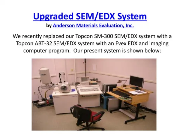
Upgraded SEM/EDX Systemby Anderson Materials Evaluation, Inc. We recently replaced our Topcon SM-300 SEM/EDX system with a Topcon ABT-32 SEM/EDX system with an Evex EDX and imaging computer program. Our present system is shown below:
We have realized a number of improvements as a result. Among these are:Image digitization at 256 x 256, 512 x 512, 1024 x 1024, 2048 x 2048, 4096 x 4096, and 8192 x 8192 pixels. The old system only digitized SEM secondary electron (SED) and backscatter (BSD) images at 1024 x 1024 pixels. Most of our SED and BSD images will now be taken at higher pixel resolution, while 512 x 512 is often the best resolution for EDX elemental mapping, since the x-ray emission volume beneath the surface is much larger than the surface beam entrance cross section. EDX samples about 1 to 2 micrometers deep into a sample and the emission volume is about 1 micrometer wide, so the resolution is much lower in EDX mode than in SED mode. • We can now map up to 8 elements at one time in the EDX mapping mode, or while making line scans. We were previously only able to perform the mapping of one element at time. This greatly reduces the time it takes to produce a map of several elements. We can now easily color code each characteristic x-ray emission map and combine them in various ways, including as superpositions on a SED surface topography image.
The EDX energy dispersive detector is on the same side of the chamber that the secondary electron detector is on, which means both modes image the sample optimally without having to rotate the sample with respect to the incoming electron beam as we had to do on the SM-300 system. Electron beam shadowing effects are now similar in both the SED and EDX images. • The EDX x-ray count rates are slightly higher with the new detector, especially at the lower mass element end of the spectrum. We are getting a slightly higher energy resolution also with this detector. • The new Robinson Back Scatter detector has a slightly higher count rate than our previous detector did. The back-scatter detector allows one to see higher mass element concentration areas as slightly brighter than lower mass elements. Because one is only detecting high energy electrons from the sample, the volume emitting the signal is substantially deeper into the sample than in a SED image. The resolution is somewhat less in the BSD mode than in the SED mode due to sub-surface spreading of the electron beam before many of the high energy electrons are back-scattered.
SED images can generally be made at higher resolution than on the SM-300 system. The resolution specification is 5 nm. The SM-300 had a specification of 4 nm, but this was impractical because achieving that resolution required such small apertures in the electron beam column that they quickly became dirty with carbon accumulations. For practical use, we therefore had to replace the really small apertures with larger apertures and our resolution was degraded to about 7 nm. The ABT-32 system is based on a very different aperture geometry which allows larger apertures placed differently. Practical operation at up to 40,000 times magnification is now reasonable. • All the data is acquired with one computer now, rather than one for SED and BSD images and another for EDX analysis. Export of data and images into reports is also more easily accomplished now. • The sample chamber is slightly larger now than it was in the SM-300, though that was also a large chamber.
We have a wider variety of sample holders and clamps. • The belt sample manipulation controls are tighter on this system. Those on the SM-300 had more hysteresis than one would like. • Dr. Lorrie A. Krebs and Dr. Kevin A. Wepasnick are the primary SEM/EDX users and both of them are very much enjoying using the improved system. Please feel free to talk to them directly about any SEM/EDX analysis projects you may have.
Contact Us: Anderson Materials Evaluation, Inc. 9051 Red Branch Road, Suite C, Columbia, MD 21045 Ph: (410) 740-8562 Toll Free: (866) 350-8882 Fax: (410) 740-8201 Email: contactus@andersonmaterials.com