Download
1 / 1
10 likes | 202 Views
AN EOSINOPHILIC GASTROENTERITIS IN A CAPTIVE CAPYBARA (HYDROCHOERUS HYDROCHAERIS) K ivilcim Sonmez * , G ulbin Sennazli , A ydin Gurel Istanbul University V eterinary Medicine Faculty Pathology Department TURKEY. ISTANBUL UNIVERSITY. HISTORY

E N D
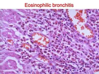
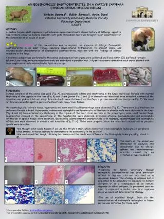
AN EOSINOPHILIC GASTROENTERITIS IN A CAPTIVE CAPYBARA (HYDROCHOERUS HYDROCHAERIS) KivilcimSonmez*, GulbinSennazli, AydinGurel Istanbul UniversityVeterinary MedicineFaculty PathologyDepartment TURKEY ISTANBUL UNIVERSITY HISTORY A captive female adult capybara (Hydrochoerushydrochaeris) with clinical history of lethargy,appetiteloss,tremors, singultus, balance disorder, soft gaitaand sudden death was brought to ourDepartmentforthedetermination of cause of death. The purpose of this presentationwas to register the presence of Allergic Eosinophilic gastroenteritis in an adult female capybara (Hydrocherushydrocheris), to present macro and microscopically characteristics of Eosinophilic gastroenteritis, together with the findings of allergic reactions in the lungs. A systemic autopsy was performed totheanimaland samples from organs were collected and fixed within10% buffered formalin solution. Later they were processed routinely and embedded in paraffin wax.3-4µ sections were taken from each organ, stained with hematoxylin-eosin and examined under light microscope. FINDINGS General condition of theanimalwasgood (Fig. A). Macroscopically edema and emphysema in the lungs, multifocal fibrosis with marked thickening of the capsule in theliver (Fig. B)and ulcers (arrowFig. C and D) in stomach and intestines were detected. Content of the intestines was pink-reddish and sassy. Intestinal walls were thickened and the Peyer’s patches were distinctive (arrowFig. E).Wecould not findanyparasiticagent in gastrointestinaltrack, lung / livertissues. Histopathologically; in brain tissue, hyperaemia and some small focal haemorrhage were observed (Fig. F). There were local hyalinisation and some fibrosis in heart. Hyperaemia, oedema and eosinophilic and lymphocytic infiltrations in alveolar walls were observed in the lungs (Fig. G and H). Non-inflammatory thickening in the capsule of the liver, focal necrosis in the parenchyma, some small haemorrhage, degenerative changes in the parenchyma of the hepatocytes were observed. Lymphoid atrophy, haemosiderosis and eosinophilic infiltration in spleen tissue were observed. Eosinophilic gastroenteritis characterized with necrosis, haemorrhages and eosinophilic infiltrations were observed in stomach and intestines (Fig. I- K). Slides were prepared from the intestinal content and no parasitic agents were detected. A B D C E The death of the animal was attributed to asphyxia and dehydration due to allergic asthma and necrohemoragic enteritis caused by an irritation or hyper sensibility -WethoughtwhatwouldhappenifweusetheWright’sstain, whichdistintivelystaineosinophileleukocytes in peripheralbloodsmears, in tissuesectionstodemonstratetheeosinophilly in thesection? -Weused a modified Wright StainingfortissuesandtheresultwasdifferentialforEosinophileleukocytes(Fig. H and L- O). RESULTS -AccordingtoMerckVeterinary Manual eosinophilicgastroenteritishas been previously defined in dog and catsanddescribed as; a uncommon disease. The etiology is unknown but is suspected to be secondary to exposure to dietary allergens or as a consequence of an immune response to other antigens.Inpresentedcasewetriedtorepresent a similarcase in a capybarawhichwecould not determinetheetiology. -Theuse of modified Wright Stainforthedemonstration of eosinophileleukocytes in tissuesectionwasdefinitiveforthesecells. H F G J I K *Corresponding Author: kivilcim@istanbul.edu.tr ThispresentationwassupportedbyIstanbul University Scientific Research Projects (Project number: 35770) L M O