Radiation Protection Update
760 likes | 1.24k Views
Radiation Protection Update. Pennsylvania Dental Association Revised December 2013 Reviewed by the Pennsylvania Department of Environmental Protection. Radiation Protection Update. Radiation in the Healing Arts is an invaluable tool with many obvious benefits.

Radiation Protection Update
E N D
Presentation Transcript
Radiation Protection Update Pennsylvania Dental Association Revised December 2013 Reviewed by the Pennsylvania Department of Environmental Protection
Radiation Protection Update Radiation in the Healing Arts is an invaluable tool with many obvious benefits. Although greatly beneficial, radiation can pose serious health risks to patients and equipment operators if not used properly. In addition to risks caused by improper practices, Americans face a much higher risk of developing cancer due to overuse of radiation for medical testing.
Radiation Protection Update The critical importance of safe and effective use of radiation in the Healing Arts, while reducing risks to patients and equipment operators, has resulted in government oversight/regulation, including regulation of dental offices and personnel. Oversight/regulation by government is handled by agencies administering and enforcing laws and regulations.
Radiation Protection Update • In Pennsylvania, the two regulatory agencies are: • The Department of State • The Department of Environmental Protection • The two governing statutes are: • The Dental Law, 63 P.S. § 120, et seq. • Radiation Protection Act, 35 P.S. § 7110.101, et seq.
Radiation Protection Update • Pennsylvania Department of State • Administers and enforces The Dental Law, which provides for initial licensure and certification requirements for dental professionals • Handled through the Department of State, Bureau of Professional and Occupational Affairs
Radiation Protection Update • Pennsylvania Department of Environmental Protection (DEP) • Administers and enforces the Radiation Protection Act (aka the “X-Ray Law”), which provides for equipment registration and inspections, and ongoing training requirements for personnel operating X-ray equipment • Handled through the Department’s Bureau of Radiation Protection
Radiation Protection Update In addition to administering and enforcing the two governing statutes, the Department of State and the DEP both have regulations to implement these statutes. Regulations, like statutes, have the force and effect of law. Policies, or “Technical Guidance” (the term used by the DEP), also guide agency action and interpretation of law, but do not themselves have force of law.
Radiation Protection Update Nearly all regulatory issues concerning X-rays in dental offices are addressed by DEP under the Radiation Protection Act (RPA), which became law in 1984. RPA governs “radiation sources,” such as X-ray equipment; and “radiation source users,” which includes any person who owns or is responsible for a radiation source. RPA gives broad regulatory powers to DEP.
Radiation Protection Act Regulation of X-rays in dental offices is handled under DEP’s regulations entitled “X-RAYS IN THE HEALING ARTS”. Regulations are found Pennsylvania’s Administrative Code, 25 Pa. Code Chapter 221. In 1998, additional regulations were promulgated which included an “Appendix A” to address training and competency for individuals who operate X-ray equipment.
Radiation Protection Act Chapter 221 requires verification of initial competence and ongoing continuing education. Determination of initial competence is based on 11 factors.
Determination of Competence • Individuals shall be trained and competent in the general operation of the X-ray equipment, and in the following subject areas: • Basic properties of radiation • Units of measurement • Sources of radiation exposure • Methods of radiation protection • Biological effects of radiation exposure • X-ray equipment • Imaging and processing • Patient exposure and positioning • Procedures • Quality assurance program • Regulations
Initial Competency Verification Any individual who satisfies the requirement of the Department of State Professional and Occupational Standards to practice in his or her discipline is deemed by DEP to have met the initial competency requirement. DEP recognizes that the Department of State determines initial competency requirements not only for physicians/dentists, but “auxiliary personnel” as well.
Continuing Education In addition to the initial determination of competence, the regulations further state that, “there shall be continuing education in radiation safety, biological effects of radiation, quality assurance and quality control.”
I. Radiation Safety Written safety procedures and rules shall be available at all facilities, including descriptions of the operating technique required for the safe operation of the particular X-ray system. The operator shall be familiar with the rules.
I. Radiation Safety Review patient’s medical history and physical condition prior to taking X-rays. Be willing to accommodate needs of the patient. Verify non-pregnancy of female patients prior to taking X-rays. Postpone elective x-rays until pregnancy is over. Inquire about any past radiation therapy. If a patient has a history of large radiation exposure over the last two years, elective radiographs should be minimized.
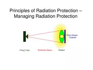
I. Radiation Safety All ionizing radiation is harmful. To protect the patient and the operator from excess exposure, radiation exposure guidelines have been established. The amount of radiation that reaches the body of the operator can be measured through use of a monitoring device known as a film badge.
I. Radiation Safety Radiation is measured in units called the sievert or gray (formerly known as rem or rad). Maximum permissible dose (“MPD”) recommendations specify the dose limits for exposure to the ionizing radiation without tissue damage. The MPD for those who work with radiation is 0.05 sievert/year (System International – SI Units) or 5.0 rems/year. For the public, it is 0.001 Sv/year or 0.1 rem/year. The MPD for operators who are pregnant is 0.001 Sv/year or 0.1 rem/year, for the entire pregnancy.
I. Radiation Safety • Effective Dose (Sv) The effective dose is used to estimate risk in humans. It allows the risk from exposure to one region of the body to be compared with the risk from exposure to another region. It also considers the radio sensitivities of different tissues for cancer formation or heritable effect.
Summary of Radiation Quantities and Units**Data from The NIST reference on Constants, Units and Uncertainty
I. Radiation SafetyKilovoltage A kilovolt is a measure of electrical current and is equal to 1000 Volts. The higher the voltage, the more energetic (thus a higher penetrating potential) the X-ray will be. Lower energy X-rays are absorbed by the body and objects through which they pass. X-rays that are absorbed do not make it to the film to create an image.
I. Radiation SafetyKilovoltage Kilovoltage peak (kVp) is the maximum amount of voltage used in an X-ray machine. Some machines can be adjusted from 50 to 100 kVp. Other machines are preset to 70 kVp. Settings below 60 and above 85 kVp are rarely used in dentistry.
I. Radiation SafetyKilovoltage • Every 15% increase in kV, will cause a doubling in the exposure or radiographic density. • Going from 70 to 80 kVp would double the exposure. To compensate, the exposure time would have to be decreased by half. • Going from 70 to 60 kV would decrease the exposure by half. To compensate, the exposure time would need to be doubled.
I. Radiation SafetyMilliamperage A milliampere (mA) is measure of electrical current and is equal to 1/1000A The higher the amperage, the higher quantity of X-rays there will be. Some machines can be adjusted from 10 to 15 mA. Some machines are preset to 6 or 7 mA.
I. Radiation Safety Utilization of proper equipment aids in limiting the amount of radiation dental patients receive. The dental X-ray tubehead must be equipped with appropriate aluminum filters, lead collimator, and position-indicating device. State and federal laws regulate the required thickness of the aluminum filters. Machines that operate below 70 kVp require a minimum of 1.5mm aluminum thickness if manufactured after December 1, 1980. Machines manufactured before December 1, 1980 require a minimum of 0.3mm if operating below 51 kVp and a minimum of 1.2mm if operating between 51 kvp – 70 kVp. All Machines operating above 70 kVp require a minimum of 2.1mm aluminum thickness. Collimation is used to restrict the size and shape of the X-ray beam. It is a lead plate with a hole in the middle and is fitted over the opening of the machine housing where the X-ray beam exits the tubehead.
I. Radiation Safety • The position-indicating device (PID) appears as an extension of the X-ray tube and is used to direct the X-ray beam. • There are three types of PIDs: • Conical (no longer used in dentistry) • Rectangular • Round
I. Radiation Safety Rectangular and round PIDs are available in varying lengths from short (8”) to long (16”). The long PID is preferred. The rectangular type is the most effective in reducing patient exposure and can reduce patient dose by 60 %. Thyroid collars, lead aprons, fast film, and film-holding devices all reduce radiation received by the patient.
I. Radiation Safety The lead thyroid collar protects the thyroid gland from scatter radiation. The lead prevents radiation from reaching the highly radiosensitive tissues of the thyroid. The thyroid collar may be separate or connected to the lead apron. The thyroid collar is recommended for all intraoral films. It is not recommended for extraoral films because it obscures information on the film (example – panorex).
I. Radiation Safety • Intensifying Screens and Film - Extraoral films are typically screen films and require the use of intensifying screens and a cassette for exposure - The two most common extraoral films include panoramic and cephalometric films - Rare earth intensifying screens are recommended combined with high speed film of 400 or greater. (ADA 2006) - Rare earth screens decrease patient exposure by as much as 55% in panoramic and cephalometric radiology.
I. Radiation Safety • Source to Skin Distance Use of a long source-to-skin distance of 40cm rather than shorter distances of 20cm, decreases exposure by 10 to 25%. Distances between 20cm and 40cm are appropriate, but the longer distances are optimal. (ADA 2006)
I. Radiation Safety Fast film is the most effective method of reducing a patient’s exposure to radiation. Fast film is available for intraoral and extraoral radiography. F-speed is the fastest intraoral film currently available. It offers a 60% reduction over D-speed film. Do not use film slower than D-speed. Current digital sensors offer greater or equal dose savings.
I. Radiation Safety The dentist must prescribe X-rays based on individual needs. The dentist uses professional judgment to determine the number, type, and frequency of the dental radiographs. The ADA, in conjunction with the FDA, has adopted guidelines for prescribing X-rays.
I. Radiation Safety There are three guiding principles in radiation protection: 1) principle of justification 2) principle of optimization 3) principle of dose limitation
I. Radiation Safety Principle of Justification - obligates the dentist to do more good than harm - dentist should identify situations where the benefit to the patient from diagnostic exposure exceeds the low risk of harm
I. Radiation Safety Principle of Optimization - holds that dentists should use every means to reduce unnecessary exposure to their patients and themselves - This philosophy of protection is known as the principle of ALARA (As Low As Reasonably Achievable).
I. Radiation Safety Principle of Dose Limitation - Dose limits are used for occupational and public exposures to ensure that no individuals are exposed to unacceptable high doses.
I. Radiation SafetyMinimizing Radiation Use fastest film possible Expose as few films as possible Use correct exposure values Use film aiming devices Process films correctly Use appropriate filters Consult doctor if film is flawed or has artifact
I. Radiation SafetyOperator • The dental radiographer must use proper protection measures to avoid occupational exposure. • Proper procedures include: • Avoid the primary beam • Stand at least 6 feet away from the X-ray tubehead • Stand in the safe zone which is 90-135 degrees from the direction of the primary beam • Never hold a film in place during exposure • Never hold the tubehead during exposure
II. Biological Effects of Radiation All ionizing radiations can produce biologic changes in living tissues and must be used judiciously. Although the amount of X-radiation used in dentistry is very small, biologic damage can still occur.
II. Biological Effects of Radiation Not all X-rays pass through the patient to reach the dental X-ray film. Some X-rays are absorbed by the patient’s tissues. Chemical changes can occur that result in biologic damage.
II. Biological Effects of Radiation There are two types of radiation injury that can occur: 1) Ionization 2) Free radical formation
II. Biological Effects of Radiation Ionization • May occur when X-rays strike a patient’s tissues • Causes formation of a positive atom and a dislodged negative electron • The excited electron can cause a chemical change in the cell that results in biologic damage • Ionization has little effect if the chemical changes do not alter sensitive molecules • Can cause substantial changes in DNA
II. Biological Effects of Radiation Free Radical Formation • X-radiation may cause the formation of free radicals, which are the primary cause of cell damage • Highly reactive and unstable atom • May recombine with other free radicals to cause cell damage • May combine with ordinary molecules to form a toxin
II. Biological Effects of Radiation Short Term Effects– large amounts of radiation delivered in minutes, days, or weeks. Not applicable to dentistry. Long Term Effects – small amounts of radiation absorbed repeatedly over a long period of time.
II. Biological Effects of Radiation Patient Exposure and Dose • Patient dose from dental radiography is usually reported as the amount of radiation received by a target organ. Although actual exposures vary considerably, dental exposures are a small fraction of the annual average background exposure. • The most radiosensitive target organs commonly studied include bone marrow, thyroid gland, brain, and salivary glands.
II. Biological Effects of Radiation Sources of Ionizing Radiation 1) Natural (background) 2) Technologically induced
II. Biological Effects of Radiation Annual Estimated Average Effective Dose Equivalent Received by a Member of the Population of the US SourceAverage Annual Effective Dose Equivalent (Sv) (mrem) Inhaled (Radon and Decay Products) 2000 200 Other Internally Deposited Radionuclides 390 39 Terrestrial Radiation 280 28 Cosmic Radiation 270 27 Cosmogenic Radioactivity 10 1 Rounded Total from Natural Sources 3000 300 Rounded Total from Artificial Sources 600 60 Total 3600 360 *Source: University of Michigan11
II. Biological Effects of Radiation As shown in the table on the previous slide, 82% of the total average annual effective dose is from natural sources of radiation, and of that, most is from radon. Of the other 18%, the majority is from medical diagnosis and treatments, with less than one percent from nuclear power and fallout.
II. Biological Effects of Radiation Effective Dose From Diagnostic X-Ray Examinations Dental ExaminationsEffective dose (mSv) -Rectangular collimation Posterior bitewings: PSP or F-speed film .005 FMX: PSP or F-speed film .035 -Round collimation FMX: PSP or F-speed film .171 FMX: D-speed film .388 -Panoramic .009 - .026 -Cephalometric .003 - .006 -Cone-beam Imaging .058 - .206 **occupational dose limit – 50 mSv per year **non-occupational (public) dose limit -1 mSv per year *Source: White and Pharoah4
II. Biological Effects of Radiation Effective Dose From Diagnostic X-Ray Examinations Medical ExaminationsEffective dose (mSv) Chest (2 views) 0.1 Cervical spine 0.20 Thoracic spine 1.0 ‘ Mammography 0.4 Extremity 0.001 Upper GI 6.0 Barium enema 8.0 CT scan – head 2.0 CT scan – chest 7.0 *Source: Mettler and Upton2
II. Biological Effects of Radiation Somatic effects - Are seen in the person irradiated • Are not transferred to future generations • Radiation injuries can produce changes in somatic cells and result in poor health • Somatic cells include all cells except reproductive