Download
1 / 1
10 likes | 190 Views
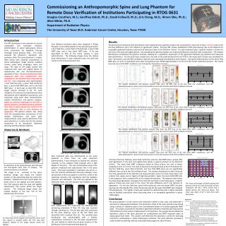
Commissioning an Anthropomorphic Spine and Lung Phantom for Remote Dose Verification of Institutions Participating in RTOG 0631 Douglas Caruthers, M.S.; Geoffrey Ibbott, Ph.D.; David Followill, Ph.D.; Eric Chang, M.D.; Almon Shiu, Ph.D.; Allen White, Ph.D. Department of Radiation Physics

E N D
Commissioning an Anthropomorphic Spine and Lung Phantom for Remote Dose Verification of Institutions Participating in RTOG 0631 Douglas Caruthers, M.S.; Geoffrey Ibbott, Ph.D.; David Followill, Ph.D.; Eric Chang, M.D.; Almon Shiu, Ph.D.; Allen White, Ph.D. Department of Radiation Physics The University of Texas M.D. Anderson Cancer Center, Houston, Texas 77030 Introduction The RPC developed a new phantom to ensure comparable and consistent radiation administration in spinal radiosurgery clinical trials, particularly the Radiation Therapy Oncology Group protocol 0631. This study assessed the phantom’s dosimetric and anatomic utility. The ‘spine phantom’ is a water filled thorax with anatomy encountered in spinal radiosurgery: target volume, vertebral column, spinal canal, esophagus, heart, and lungs. The dose to the target volume was measured with axial and sagittal planes of radiochromic film and thermoluminescent dosimeters (TLD). The dose distributions were measured with the radiochromic film calibrated to the absolute dose measured by the TLD. Four irradiations were administered: a four field box plan, a seven field conformal plan, a seven field IMRT plan, and a nine field IMRT plan. In each plan, at least 95% of the target volume received 8 Gy. For each irradiation the planned and administered dose distributions were registered via pinpricks, and compared using point dose measurements, isodose distributions, and gamma analyses. Based on previous experience at the RPC, a gamma analysis is considered passing if greater than 95% of pixels passed the criteria of 5% dose difference and 3 mm distance to agreement. This gamma analysis test, along with the complementary assessments of the isodose distributions and point dose measurements, were used to determine if the spine phantom is a useful tool for the remote assessment of an institution’s treatment planning and dose delivery regimen. Results Four different treatment plans were designed in Philips Pinnacle 7.6 and administered to the spine/lung phantom: a four field box, a seven field conformal plan, a seven field IMRT plan, and a nine beam IMRT plan. 8 Gy was prescribed to 95% of the tumor volume in each administration. The following images show the relative dose distributions in each treatment plan; the dark blue contour is the 8 Gy prescription line. The planned and measured dose distributions were registered and a gamma analysis was conducted for each trial of each treatment plan at the 5% dose difference and 3 mm distance to agreement criteria. Previous RPC studies established a 95% pixel passing rate as the baseline for acceptable agreement between planned and measured dose at the 5%/3mm criteria. For each treatment plan, a gamma analysis is shown for one trial in the axial and sagittal planes. Accompanying the gamma analyses are isodose distributions and point dose comparisons. The isodose distributions complement the gamma analyses, allowing for a qualitative assessment of the agreement between the planned and measured dose distributions. For the point dose comparisons, the ratio of the planned dose (from the TPS) to the measured TLD dose was calculated for each TLD location, and the 95% confidence intervals were calculated and plotted at each location. During the administration of the seven field IMRT plan, an error in localization was made; the phantom was shifted approximately 2.5 mm from the correct treatment position. The results from the seven field IMRT irradiations are included for illustrative purposes. The isodose distribution and gamma analysis map from the first trial of the four field box irradiations, and the planned/measured ratios at each TLD location (n=3 at each location) with 95% confidence intervals. Four field box plan dose distribution Seven field conformal plan dose distribution The isodose distribution and gamma analysis map from the first trial of the seven field conformal irradiations, and the planned/measured ratios at each TLD location (n=3 at each location) with 95% confidence intervals. Materials & Methods The isodose distribution and gamma analysis map from the first trial of the seven field IMRT irradiations, and the planned/measured ratios at each TLD location (n=3 at each location) with 95% confidence intervals. Seven field IMRT plan dose distribution The isodose distribution and gamma analysis map from the first trial of the nine field IMRT irradiations, and the planned/measured ratios at each TLD location (n=3 at each location) with 95% confidence intervals. Nine field IMRT plan dose distribution Each treatment plan was administered to the spine phantom in three trials. For each treatment administration, it was necessary to localize the physical isocenter to the radiation field isocenter with a high degree of accuracy. This was accomplished using cross patterns irradiated onto radiochromic film in the spine insert. The linear accelerator’s collimator was set to a 1 mm ‘slit’, and the radiochromic film was irradiated. From the position of the cross pattern on the film, a shift of the treatment isocenter into coincidence with the radiation isocenter was calculated. The shift was applied to the treatment table, and this process was iterated until full coincidence was achieved. All trials of the four field box, seven field conformal, and nine field IMRT plans exceed 95% pixel agreement in the axial and sagittal dose planes in gamma analysis at the 5%/3mm criteria. The seven field IMRT irradiations failed at these criteria, but with a known localization error of 2.5 mm. The ratio of planned to measured dose at the TLD locations for the four field box, seven field conformal, and nine field IMRT plans are not significantly different from unity at the 5% confidence level. The isodose distributions further illustrate the close agreement of the planned and measured dose across for these three plans; the isodose lines agree, even in regions of high dose gradient. Again, the seven field IMRT plan is the exception, with a notable shift on the isodose distributions. The adjacent chart shows the mean pixel agreement (average of the trials) for each plan in each dose plane. A tighter gamma criteria was also used in this chart: 3% dose difference and 2 mm distance to agreement. For the four field box, seven field conformal, and nine beam IMRT, the pixel passing rate still exceeds 95%, while the passing rate for the seven field IMRT plan dropped off precipitously. This tighter criteria was explored to demonstrate that a higher standard may be feasible, particularly when discussing what is an acceptable agreement in spinal radiosurgery. An illustration of the spinal phantom with mid-axial and mid-sagittal views from CT imaging. The anatomical features are labeled. This image is an overview of the spine phantom design, and shows the relative location of the anatomical features within the phantom. Radiochromic film inserts bisect the spinal insert in the sagittal and axial aspects. Four TLD capsules abut the axial and sagittal radiochromic film inserts within the target volume. The following image shows the relative location of the four TLD to the radiochromic film inserts. The mean percentage of pixels passing the gamma criteria for each dose plane of each irradiation; the 5% / 3mm criteria and a tighter criteria, 3% / 2mm, are shown. The mean was taken of all 3 trials from each dose plane. Conclusion The spine phantom is to be used to test institution’s ability to scan, plan, and administer a stereotactic radiosurgical treatment. The dosimetric utility of the spine phantom was tested using a variety of irradiation plans, from unmodulated beams to clinically applicable IMRT plans. This project assessed the dosimetric agreement between treatment planning and the TLD/radiochromic film system implemented in the phantom. This project demonstrated the dosimetric utility of the spine phantom for unmodulated and IMRT treatment plans at radiosurgical dose levels. This project confirmed that the phantom is useful for assessing institutions participating in spinal radiosurgery protocols. The spine phantom provided a useful model for planning intensity modulated radiosurgery for spinal tumors. References Davidson, S. E. (2006). Heterogeneity Dose Calculation Algorithm Accuracy in IMRT Using Anthropomophic Thorax Phantom. Houston, The Graduate School of Biomedical Sciences. Low, D. A., W. B. Harms, S. Mutic and J. A. Purdy (1998). "A technique for the quantitative evaluation of dose distributions." Med Phys25(5): 656-61. Molineu, A., D. S. Followill, P. A. Balter, W. F. Hanson, M. T. Gillin, M. S. Huq, A. Eisbruch and G. S. Ibbott (2005). "Design and implementation of an anthropomorphic quality assurance phantom for intensity-modulated radiation therapy for the Radiation Therapy Oncology Group." Int J Radiat Oncol Biol Phys63(2): 577-83. RTOG (2009). RTOG 0631 Phase II/III Study of Image-Guided Radiosurgery/SBRT for Localized Spine Metastasis. http://www.rtog.org/members/protocols/0631/0631.pdf. 1) 2) 3) 4) Each radiochromic film underwent the following data processing procedure: A Raw OD map was acquired through scanning (1). The OD data was then processed by the dose response curve of the film batch and smoothed with a median filter (2). The resultant dose distribution was downsampled with a bilinear interpolation, corrected to the TLD dose measurement (3), and rotated to match the pin-prick locations of the distribution from the planned dose plane (4). An illustration of the sagittal and axial film slices; each TLD capsule is located within the PTV, and their locations relative to the target volume center are labeled. The investigation was supported by PHS grants CA10953 and CA81647 awarded by the NCI, DHHS.


