Chapter 24
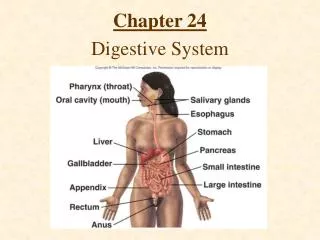
Chapter 24. Digestive System. Digestive System Anatomy. Digestive tract Alimentary tract or canal GI tract Accessory organs Primarily glands Regions Mouth or oral cavity Pharynx Esophagus Stomach Small intestine Large intestine Anus. The function of digestive system:

Chapter 24
E N D
Presentation Transcript
Chapter 24 Digestive System
Digestive System Anatomy • Digestive tract • Alimentary tract or canal • GI tract • Accessory organs • Primarily glands • Regions • Mouth or oral cavity • Pharynx • Esophagus • Stomach • Small intestine • Large intestine • Anus
The function of digestive system: • Mechanically breakdown the food stuff and enzymatically digest them. • Absorb necessary nutrients including ions. • Lubrication, emulsification, mixing, transport - Deglutition, bolus, peristalisis, etc. • Release of wastes from the body
Functions • Ingestion: Introduction of food into stomach • Mastication: Chewing • Propulsion • Deglutition: Swallowing • Peristalsis: Moves material through digestive tract
Functions • Mixing: Segmental contraction that occurs in small intestine • Secretion: Lubricate, liquefy, digest • Digestion: Mechanical and chemical • Absorption: Movement from tract into circulation or lymph • Elimination: Waste products removed from body
Nervous regulation Involves enteric nervous system Types of neurons: sensory, motor, interneurons Coordinates peristalsis and regulates local reflexes Chemical regulation Production of hormones Gastrin, secretin Production of paracrine chemicals Histamine Help local reflexes in ENS control digestive environments as pH levels Digestive System Regulation
Oral Cavity • Mouth or oral cavity • Vestibule: Space between lips or cheeks and alveolar processes • Oral cavity proper • Lips (labia) and cheeks • Palate: Oral cavity roof • Hard and soft • Palatine tonsils • Tongue: Involved in speech, taste, mastication, swallowing
Salivary Glands • Produce saliva • Prevents bacterial infection • Lubrication • Contains salivary amylase • Breaks down starch • Three pairs • Parotid: Largest • Submandibular • Sublingual: Smallest
a. Functions of the oral cavity • In addition to chewing, or mastication in the oral cavity its enzyme contributes the initial stage of digestion.The salivary glands release salivary amylase, which digests starch into glucose and the other polysaccharides to disaccharides. (Fig. 24-7 and Fig. 24.9)Note that humans do not digest cellulose, since they do not have cellulase.In addition, saliva contains lysozyme and immunoglobulin A to fight against bacteria.Mucous release from the other glands in the oral cavity contains mucin, a proteoglycan, that contributes lubrication of the mouth.The salivary glands stimulation is primarily through the parasympathetic nerve.Higher centers of the brain also affect the activity of the salivary glands.
Pharynx Nasopharynx Oropharynx: Transmits food normally Laryngopharynx: Transmits food normally Esophagus Transports food from pharynx to stomach Passes through esophageal hiatus (opening) of diaphragm and ends at stomach Hiatal hernia Sphincters Upper Lower Pharynx and Esophagus
Deglutition (Swallowing) • Three phases • Voluntary • Bolus of food moved by tongue from oral cavity to pharynx • Pharyngeal Reflex: Upper esophageal sphincter relaxes, elevated pharynx opens the esophagus, food pushed into esophagus • Esophageal • Reflex: Epiglottis is tipped posteriorly, larynx elevated to prevent food from passing into larynx
Stomach Anatomy • Openings • Gastroesophageal: To esophagus • Pyloric: To duodenum • Regions • Cardiac • Fundus • Body • Pyloric
Stomach Histology • Layers • Serosa or visceral peritoneum: Outermost • Muscularis: Three layers • Outer longitudinal • Middle circular • Inner oblique • Submucosa • Mucosa
Stomach Histology • Rugae: Folds in stomach when empty • Gastric pits: Openings for gastric glands • Contain cells • Surface mucous: Mucus • Mucous neck: Mucus • Parietal: Hydrochloric acid and intrinsic factor • Chief: Pepsinogen • Endocrine: Regulatory hormones
i. Secretions in the stomach (Fig. 24-13) • The mixture in the stomach is called chyme.Stomach secretes mucus, hydrochloric acid, gastrin, intrinsic factor, and pepsinogen, a precursor to protease pepsin.Alkaline mucus secreted from the mucus cells protects the epithelial cells from the acidic chyme and pepsin. Parietal cells in the gastric glands secrete intrinsic factor and concentrated HCl. Intrinsic factor is a vitamin B12 binding glycoprotein for better absorption of B12. • Chief cells within the gastric glands secrete pepsinogen, which will be activated to pepsin by HCl.
i. Low pH in the stomach • Relatively high concentration of HCl released in the stomach has the following functions • (a) The value of pH in the stomach is 1 - 3. • (b) Most of bacteria are killed at this pH. But not all! - Pylori • (c) Inactivate amylase, thus no further digestion of carbohydrates. • (d) Many proteins are denatured. • (e) Pepsin, now activated at this pH, can digest these proteins
i. Secretion of H+ • Release of H+ from parietal cell into the stomach starts with consumption of CO2 from blood. (Fig. 24-14) Note the names of players in this process: CA, ATP requiring proton/potassium exchange pump, bicarbonate/chloride shift, movements of potassium, bicarbonate and chloride ions.
i. Regulation of stomach secretion • 2 - 3 L of gastric secretion/day. • Up to 700 ml/meal depending on the types of meals. • Both neuronal and hormonal regulations are possible. For details study Fig. 24-15.
Small Intestine • Site of greatest amount of digestion and absorption • Divisions • Duodenum • Jejunum • Ileum: Peyer’s patches or lymph nodules • Modifications • Circular folds or plicae circulares, villi, lacteal, microvilli • Cells of mucosa • Absorptive, goblet, granular, endocrine
Small Intestine Secretions • Mucus • Protects against digestive enzymes and stomach acids • Digestive enzymes • Disaccharidases: Break down disaccharides to monosaccharides • Peptidases: Hydrolyze peptide bonds • Nucleases: Break down nucleic acids • Duodenal glands • Stimulated by vagus nerve, secretin, chemical or tactile irritation of duodenal mucosa
The primary center for digestion and adsorption. In three parts: duodenum, jejunum and ileum making up to 6 meters (Fig. 24-16)In fact, the digestive function in the small intestine follows the food stuff being digested by pancreatic juice to relatively smaller molecules. Observe the anatomy and histology of the intestinal wall to find intestinal glands, capillary networks in the villi, lacteal, goblet cells, etc. (Fig. 24-17)
i. Secretions in the small intestine (Fig. 24-16, 17) • Duodenal glands and Goblet cells release mucus. • The final stage of break down to small molecules so that they may be absorbed through villi. • Absorptive cells release digestive enzymes, such as aminopeptidase, peptidase, enterokinase (trypsinogen activator), amylase, sucrase, maltase, isomaltase, lactase and lipase. • These enzymes are bound to the membranes of the absorptive cell Microville. • Many other digestive enzymes are supplied from the pancreas.
i. Movement in the small intestine • Segmental contraction for mixing and peristaltic contractions for propelling are observed. • The contractions move at rate of 1 cm/min, thus taking 3 - 5 hours for chyme to move from the pylorus to the ileocecal junction. • ii. Absorption from the small intestine • Out of about 9 L of water enters the digestive system, the small intestine absorbs about 8 - 8.5L by osmosis.
Pancreas • Anatomy • Endocrine • Pancreatic islets produce insulin and glucagon • Exocrine • Acini produce digestive enzymes • Regions: Head, body, tail • Secretions • Pancreatic juice (exocrine) • Trypsin • Chymotrypsin • Carboxypeptidase • Pancreatic amylase • Pancreatic lipases • Enzymes that reduce DNA and ribonucleic acid
a. Functions of the pancreas The gross anatomy and cytology of the pancreas is shown in (Fig. 24-18). The pancreas has both endocrine and exocrine cells.Endocrine: Pancreatic islet contact to blood stream. Alpha cells – glucagons Beta cells – insulinExocrine: Acini cells open to ducts and secrete enzymes
i. Pancreatic juice to adjust pH.The columnar epithelial cells of the pancreas contains bicarbonate ions, which will be released into the intralobular duct of the pancreas. The bicarbonate ion neutralize the acidic chyme and stops pepsin activity, while make it possible for the pancreatic enzymes to remain active in the small intestine. (Fig. 24.22 of Seeley)
i. Pancreatic enzymesThe acinar cells of the pancreas produce pancreatic enzymes.Many proteolytic enzymes are released in the form of precursors.They include, trypsinogen , chymotrypsinogen, and procarboxypeptidase and are activated by other enzymes, such as enterokinase.Amylase, lipases are also present.
i. Control of pancreatic secretionBy both hormonal and neuronal means. (Fig. 24-22 and Fig. 24.23 of Seeley)Also study Table 24-1.
Liver • Lobes • Major: Left and right • Minor: Caudate and quadrate • Ducts • Common hepatic • Cystic • From gallbladder • Common bile • Joins pancreatic duct at hepatopancreatic ampulla
Functions of the Liver • Bile production • Salts emulsify fats, contain pigments as bilirubin • Storage • Glycogen, fat, vitamins, copper and iron • Nutrient interconversion • Detoxification • Hepatocytes remove ammonia and convert to urea • Phagocytosis • Kupffer cells phagocytize worn-out and dying red and white blood cells, some bacteria • Synthesis • Albumins, fibrinogen, globulins, heparin, clotting factors
Liver functions Anatomy and histology: (Fig. 24.19) • i. Bile production • 600 - 1000 ml/day • No digestive enzymes • Dilute and neutralize stomach acid and emulsify fats. • The pH of chyme is raised so that pancreatic enzymes can function. • Contains bilirubin from broken down hemoglobin. • Cholesterol, fast, fat-soluble hormone and lecithin are found. • The blood and bile flow: (Fig. 24.20) • Stimulates bile secretion - secretin from duodenum, by parasympathetic vegas nerve, increased blood flow in the liver, etc. (Fig. 24.21)