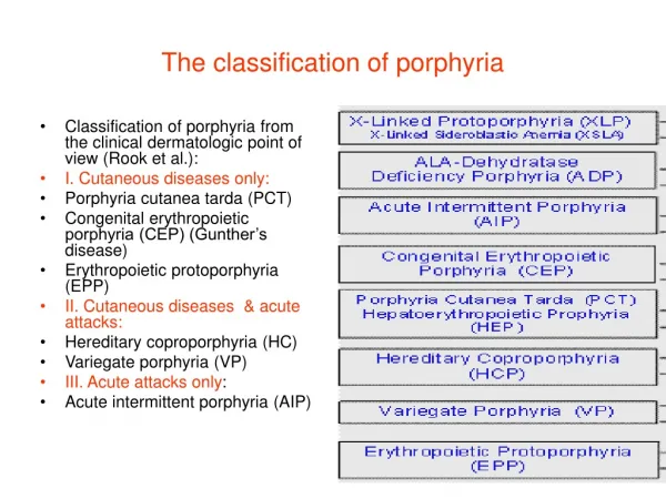
The classification of porphyria
Classification of porphyria from the clinical dermatologic point of view (Rook et al.): I. Cutaneous diseases only: Porphyria cutanea tarda (PCT) Congenital erythropoietic porphyria (CEP) (Gunther’s disease) Erythropoietic protoporphyria (EPP) II. Cutaneous diseases & acute attacks:

The classification of porphyria
E N D
Presentation Transcript
Classification of porphyria from the clinical dermatologic point of view (Rook et al.): I. Cutaneous diseases only: Porphyria cutanea tarda (PCT) Congenital erythropoietic porphyria (CEP) (Gunther’s disease) Erythropoietic protoporphyria (EPP) II. Cutaneous diseases & acute attacks: Hereditary coproporphyria (HC) Variegate porphyria (VP) III. Acute attacks only: Acute intermittent porphyria (AIP) The classification of porphyria
2O% of patients with porphyria cutanea tarda are inherited in an autosomal dominant pattern
Porphyria cutanea tardaB: urine of patient is pink under a Wood’s lamp C: Urine under ultraviolet A light. The diagnosis was confirmed by marked presence of uroporphyrinogen.Chan,C.C. & Lin,S. N. Eng. J. Med. 365:1128, 2O11
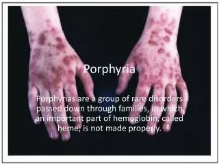
A. Blister, milia & hyper pigmented scar B. Blister, erosion & superficial scar C. Hemorrhagic blister, crusting & superficial scar D, Erosion & sclerodermiform plaque on the neck of the patient. Porphyria cutanea tarda
Porphyria cutanea tarda systemic lupus erythematosusFritsol, S. et al. Rev. Bras. Remm. 52, no. 6, 2O123
Porphyria cutanea tarda & systemic lupus erythemastosusThe use of chloroquine for treatment of SLE,may cause hepatotoxicity leading to PCTHaendcchan,L. et al.An. Bras. Dermat.86: nO.1, 2O11
Congenital erythropoietic porphyria (Gunther’s disease)It has an autosomal recessive pattern of inheritance
Erythropoietic protoporphyriaOrphanet J. Rare Disease 4 : 19, 2OO9
Erythropoietic protoporphyriaOrphanet J. Rare Disease 4 : 19, 2OO9
Hereditary coproporphyriaIt is due to mutation CPOX gene (coproporphyrinogen oxidase enzyme ) which is located on the long arm ofchromosome 3 at position 12
Porphyria variegataWhatley,S.D. al. AJHD 65:984, 1999Mutation in human PPOX gene: missense mutation ( .) are shown below the gene diagram, with chain termination& splice mutation (frameshift) (X), nonsense (open box) & splice defect (dark box) above it. Unshaded area of exons denote non coding regions
Urine is placed in a test tube A Equal amount of Echrlich’s aldehyde reaction, red color means strongly positive reaction Chloroform is added to the tube, If red color remasin in the top, this means the presence of PBG (C) which suggest acute porphyria. If the color is confined to the lower layer, the urine contain urobilinogen & this is of no significance in terms of porphyria The reaction is negative in variegate porphyria, but if it is positive, this will confirm an acute attack. Watson-Schwartz reactionScreening test for acute intermittant porphyria
AIP is caused by mutation in the HMBS gene located at 11q23.3 Acute intermittent porphyriaSequence chromatography of the PBGD (Porphobilinogen deaminase) gene demonstrate a short train of deletion( c 1OO8 – 1O19) in I-1, II-1& II-2J. of neurological science 26O: 231, 2OO7
Garlic makes the case of acute intermittent porphyria worse because it contains chemicals that excerbate the disease .(Vampire myth which means that porphyria is vampire’s disease)