Hepatic Function Analysis
Explore the complexities of liver structure, function, and pathology with a focus on metabolic and synthetic functions, enzyme-associated damage, and diagnostic tests. Understand terminologies and key markers for hepatic diseases.

Hepatic Function Analysis
E N D
Presentation Transcript
Hepatic Function Analysis Clinical Pathology January 13, 2009
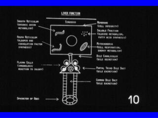
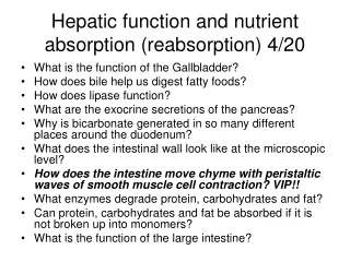
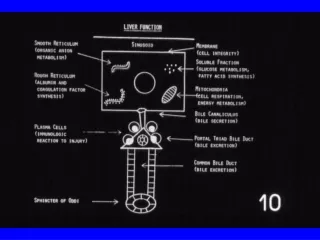
Hepatic Functions • Liver is the largest internal organ • Complex in terms of structure, function and pathology • Multitude of functions include: • Metabolic liver functions • Key role in conjugation and excretion of bilirubin and bile acids, exogenous drugs, production of proteins, metabolism of amino acids, carbohydrates and lipids and conversion of ammonia to urea. • Synthesis: albumin, cholesterol, plasma protein, clotting factors. • Digestion & Absorption: related to bile • Elimination/detoxification: toxins & catabolism of drugs.
Metabolic Functions continued • All functions are by enzymatic reactions • Malfunctions result in: • Jaundice • Hypoalbuminemia • Hemostasis problems • Hypoglycemia • Hyperlipoproteinemia • Hepatoencephalopathy
Enzymes Associated with Hepatocellular Damage • Hepatocytes are damaged and enzymes leak into the blood. • No single test is superior to any other for detecting hepatic disease although some tests are better at verifying liver function. • Hepatocytes are cuboidal to polyhedral cells and or often binucleated.
Terminology • Microhepatica: small liver, suggests PSS or fibrosis, cirrhosis. • Hepatomegaly: enlarged liver, suggests neoplasia, toxicity, systemic disease. • Hepatic Encephalopathy: Increased levels of blood ammonia causing severe behavioral changes. • Icterus: Yellow discoloration of the skin and/or serum.
Alanine Aminotransferase (ALT) • Formerly known as SGPT. • Dogs, cats, non-human primates major source is the hepatocyte. • Horses, ruminants, and pigs not liver specific. • Other sources: Renal, cardiac, skeletal muscle and pancreas. • Screening test: not precise enough to identify specific liver disease due to other sources. • Tests hepatocyte membrane integrity. • No correlation between blood levels and severity of hepatic damage. • Sample handling: avoid hemolysis & lipemia.
Alkaline Phosphatase (Alkphos or AP) • As isoenzymes in osteoblasts, chondroblasts, & hepatocellular cells • Young animals found in osteoblasts and chondroblasts due to active bone development. • Other animals most alkphos is from the liver. • Used to detect cholestasis in dogs and cats (not useful in cattle and sheep) • Levels will increase with corticosteroid use and Cushing’s • Any increase is considered significant in the cat • 3x increased levels may be normal in puppies less than 6 months of age.
Aspartate Aminotransferase (AST) • Formerly known as SGOT. • Found in mitochondrial membranes • Not liver specific, found in erythrocytes, cardiac muscle, kidneys and pancreas. • Increased blood level may indicate nonspecific liver damage. • May indicate strenuous exercise or IM injections, muscle inflammation and/or hemolysis. • If elevated, check plasma sample for hemolysis or spin hematocrit tube to check. • Hemolysis and lipemia may elevate AST (equine has much higher level tha other species)
Sorbitol Dehydrogenase (SD) • Primary source is the hepatocyte • Used in all animals • Sheep, goats and swine do not have diagnostic levels of ALT so SD is used in place. • Liver specific test for hepatocellular damage or necrosis • Disadvantages: unstable in serum & must be assayed within 12 hours of collection. • Hemolysis does not affect results.
Gamma Glutamyltranspeptidase (GGT) • Found in many tissues, primary source is the liver. • Kidneys, pancreas, intestines and muscle cells. • Elevated with liver disease, especially obstructive liver disease. • Cattle, horses, sheep & goats have higher GGT than dogs and cats. • Hemolysis has no affect on results. • Possibly more specific for hepatic disease in cats. • Associated with cholestatis.
GGT continued • Important marker of hepatobiliary disorders in large animals. • High levels are usually found in suckling neonatal young animals. • Stable, large enzyme. • On rare occasions can be used to diagnose renal tubular disease. • Reliable indicator in large animal species of damage to liver and biliary obstruction.
Albumin • Made in liver • Maintains blood volume and binds to hormones • May be lost through the gut or kidneys. • Low levels may also indicate maldigestion or malabsorption.
Liver Function Dye Excretion • BSP clearance • Sulfobromophthalein (BSP) • Dye injected IV and retrieved by the liver, conjugated and exreted in the bile. • Blood is drawn 30 minutes later • Disappearance from the plasma depends on hepatic blood flow, bile flow and hepatocellular integrity. • Horses it is used to differentiate hepatic jaundice from anorexia & hepatoencephalopathy. • Cattle for fascioliaiss, liver abscess, ketosis, and liver flukes. • Swine Aflatoxocosis • Hepatic lesions delay BSP excretion & indicate a loss of at least 55% of liver’s functional mass.
Indocyanine Green Clearance (ICG) • Used to estimate hepatic blood flow. • Poor availability of test • IV injection in horses, dogs and cats • 5-6 plasma samples at 0, 5, 10, 15, & 30 minutes post injection. • ICG concentration measured photometrically and half-life determined. • Measure clearance rate at 0, 5, 10, & 15 minutes. • Delayed in hepatic blood flow shows hepatic necrosis & bile duct obstruction.
Ammonia Tolerance • Liver disease may impair ammonia detoxification leading to hepatoencephalopathy. • Ammonium chloride orally or rectally • Used to detect abnormal portal blood flow. • Usually measured in heparinized blood sample. • May cause vomiting or CNS signs.
Prothrombin Time (PT) Prothrombin is a Vitamin K dependent coagulation factor made by the liver. Activated by tissue thromboplastin and calcium. Initiates the coagulation involving liver coagulation factors I, II, V, VII, & X but most especially VII. Indicates coagulation factor insufficiency & possible liver necrosis, cirrhosis or poor bile secretion which is necessary for Vitamin K absorption.
Bile Acid Concentrations • Synthesized by hepatocytes from cholesterol & conjugated with glycine or taurine. • Serum bile acid (SBA) elevated with any process that impairs the hepatocellular, biliar or portal enterohepatic circulation of bile acids. • Hepatic insufficiency results in an inability to extract bile acids from the blood.
Bile Acid Measurements • Bile acids are made by the liver. • The bile acids are secreted into the gut after eating. • Fast animal 12 hours, take preprandial sample, feed animal moderately fatty meal, then take postprandial blood sample 2 hours later.
Bilirubin • A metabolite of heme portion of Hemoglobin, a waste product when RBC lyses. • Liver conjugates to water soluble then converted to urobilinogen by bacterial enzymes- urobilinogen eliminated by kidneys and in the feces. • Both unconjugated and conjugated bilirubin in plasma=total bilirubin • Conjugated referred to as direct because measured directly. • Icterus: detectable in the sclera at levels 3-4 mg/dl, detectable in the serum at 1.5-2 mg/dl. Normal values are less 1.0 mg/dl for dogs and cats. • Any increase in the serum bilirubin over normal is considered significant in dogs and cats. • Horses may become icteric if anorexic.