Download
1 / 30
300 likes | 533 Views
Protein tyrosine kinases use adaptor proteins to link antigen receptors to intracellular phosphatidylinositol (PI) and MAP kinase pathways. Y. PTKs. adaptors. PI pathway. MAP kinase pathway. Regulation of gene expression through increased transcription factor activity.

E N D
Protein tyrosine kinases use adaptor proteins to link antigen receptors to intracellular phosphatidylinositol (PI) and MAP kinase pathways Y PTKs adaptors PI pathway MAP kinase pathway
Regulation of gene expression through increased transcription factor activity NFAT: nuclear localization regulated by calcium-activated phosphatase AP-1: expression and activity regulated by MAPK pathways NFkB: nuclear localization regulated by degradation
The NFAT transcription factor is normally phosphorylated and sequestered in the cytosol. The PI pathway induces calcium influx which activates Calcineurin phosphatase activity and promotes nuclear translocation of the NFAT transcription factor.
MAP kinase pathways increase AP-1 dependent transcription by increasing expression of the Fos subunit, and by increasing activity of the Jun subunit Fos -P -P
The NF-kB transcription factor is sequestered in the cytosol by IkB which is phosphorylated and degraded in response to receptor signals nucleus
Stimulated antigen receptors utilize tyrosine kinases to induce intracellular signaling pathways leading to transcription factor activation and the induction of cell growth and differentiation. Y PTKs adaptors PI pathway MAP kinase pathways Interleukin-2, and effector functions
Commitment to complete T cell activation requires many hours: MHC-peptide ligands which dissociate early can lead to partial activation No activation Partial activation time Complete T cell activation
TCR signals enhance integrin binding Low affinity MHC-peptide ligands may not engage the TCR long enough to allow enhanced integrin binding - “inside-out” signaling
Low affinity MHC-peptide ligands may not engage the TCR long enough to allow co-receptor binding and ZAP-70 activation Fast dissociation Slow dissociation Lck ZAP70
+ adhesion T cell response - adhesion t1/2 TCR:MHC-peptide Activation is dependent upon the stability of the TCR: MHC-peptide interaction, which can be regulated by adhesion molecules and co-receptor function. This may be critical for stimulation by some peptides.
Thymic maturation: what distinguishes signals which lead to negative selection, positive selection, and death by neglect? No selection - death Negative selection - death ? Thymic stromal cell CD4+8+ thymocyte Positive selection - Maturation to CD4+ or CD8+
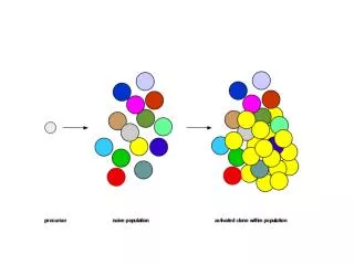
# of thymocytes TCR Affinity Positive and negative selection are a function of TCR affinity
Figure 10-13 The outcome of T helper cell differentiation can depend upon strength of TCR signal
Model of differential signaling in the thymus: the strength of signal (or time of receptor engagement) may be reflected in the extent of LAT phosphorylation and subsequent activation of MAP kinase pathways from Starr, et al. 2003, Ann. Rev. Imm 21:139
Figure 8-10 Naïve T cells are particularly dependent upon a second, co-stimulatory, signal for Interleukin-2 production leading to proliferation and also for enhanced cell survival
Figure 8-20 Induction of both Interleukin-2 (IL-2) and the high affinity IL-2 receptor stimulates T cell proliferation through an autocrine mechanism
CD28 provides 2nd signal CD28 binding to its ligand, B7, provides activation signals to the naïve T cell which are required for the production of IL-2 and increased cell survival
CD28 increases IL-2 production through increased gene transcription and through increased IL-2 mRNA stability CD28 ~~~~~~ ~~~~~~ ~~~~~~ ~~~~~~ ~~~~~~ ~~~~ IL-2
CD28 is a disulfide-linked transmembrane dimer which signals by recruiting the Vav exchange factor and PI 3-kinase to the membrane CD28 Lck Pro Pro Pro -P adaptor Vav exchange factor PI3-Kinase
Vav is an exchange factor for the Rac GTPase and stimulates one of the MAPK pathways
Like the LAT adaptor, Phosphatidylinositol 3’kinase (PI3-K) is able to induce the membrane recruitment. PI3-K produces PIP3 leading to the localized membrane binding of PH domain containing proteins. PIP2 PIP3 + ATP PH domain containing proteins PI3’Kinase Vav PLC BTK
Phosphatidylinositol 3’kinase (PI3-K) produces PIP3 leading to localized membrane binding of the Akt kinase and its activator PDK-1. Akt promotes cell survival through increased expression of BclXL and phosphorylation of BAD. PIP2 PIP3 + ATP PI3’Kinase PH domain containing proteins PDK-1 Akt ~P BclXL expression BAD phosphorylation
Figure 6-25 The mitochondrial mechanism of programmed cell death is blocked by elevated expression of anti-apoptotic Bcl2 family members (Bcl2 and BclXL).
Cell death Bcl2 function can be inhibited by interaction with the BAD protein. Phosphorylation of BAD by Akt results in its sequestration and enhanced cell survival.
CD28 costimulation enhances glucose metabolism, providing the energy stores necessary for activation K. Frauwirth et al. Immunity 16:769-777
Like CD28, CD19 on B cells also provides signals which enhance activation. CD19 function requires co-aggregation with the antigen receptor.
Figure 9-5 B cell activation is regulated by co-stimulatory signals provided by CD40. The ligand for CD40 (CD40L) is induced on activated T cells.
CD40 is a member of the TNF receptor family and activates the NF-kB transcription factor through phosphorylation of IKß kinase and subsequent phosphorylation and degradation of IKß