Download
1 / 14
140 likes | 233 Views
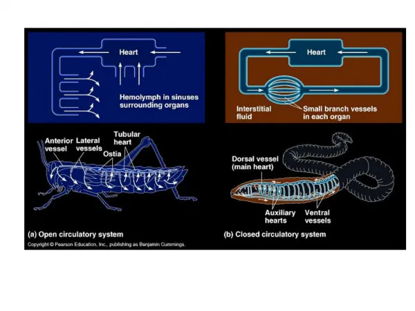
The Closed Circulatory System. Humans have a closed circulatory system , typical of all vertebrates, in which blood is confined to vessels and is distinct from the interstitial fluid. The heart pumps blood into large vessels that branch into smaller ones leading into the organs.

E N D
The Closed Circulatory System • Humans have a closed circulatory system, typical of all vertebrates, in which blood is confined to vessels and is distinct from the interstitial fluid. • The heart pumps blood into large vessels that branch into smaller ones leading into the organs. • Materials are exchanged by diffusion between the blood and the interstitial fluid bathing the cells.
The Cardiovascular System • Three Major Elements – Heart, Blood Vessels, & Blood • 1. The Heart- cardiac muscle tissue • highly interconnected cells • four chambers • Right atrium • Right ventricle • Left atrium • Left ventricle
Superior Vena Cava Right Atrium Tricuspid Valve Right Ventricle Pulmonary Semilunar Valve Lungs Pulmonary Vein Bicuspid Valve Left Ventricle Aortic Semilunar Valve Aorta To the bodies organs & cells Pathway of the blood
Circuits • Pulmonary circuit • The blood pathway between the right side of the heart, to the lungs, and back to the left side of the heart. • Systemic circuit • The pathway between the left and right sides of the heart.
Figure 19.5 Systemic & pulmonary circuits Blood from body enters Right Atrium Arteries carry blood AWAY from the heart Blood from lungs enters Left Atrium Veins carry blood to the heart (like ven in Spanish) Right Ventricle sends blood to lungs Left Ventricle sends blood to rest of body
The Cardiovascular System Blood Vessels -A network of tubes • Arteriesarterioles move away from the heart • Elastic Fibers • Circular Smooth Muscle • Not always Red (notice the pulmonary artery carries blue blood - deoxygenated blood) • Capillaries – where gas exchange takes place. • One cell thick • Serves the Respiratory System • VeinsVenules moves towards the heart • Skeletal Muscles contract to force blood back from legs • One way values • When they break - varicose veins form • Not always Blue (notice the pulmonary veins carry red blood –oxygenated blood)
Figure 19.6 Anatomical differences in the right & left ventricles
Figure 19.9A Operation of atrioventricular valves of the heart
Figure 19.9B Operation of atrioventricular valves of the heart The AV valves shown here are responsible for the Dub sound in the heart. When the valves close, they slam together and make a dub sound.