Download
1 / 28
330 likes | 607 Views
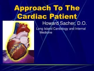
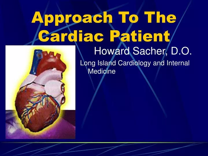
Approach To The Cardiac Patient. Howard Sacher, D.O. Long Island Cardiology and Internal Medicine. Lecture Goals and Objectives. Understand the signs and symptoms of dyspnea, chest pain, palpitations, presyncope/syncope, and fatigue Know the NYHA classification of heart dis.

E N D
Approach To The Cardiac Patient Howard Sacher, D.O. Long Island Cardiology and Internal Medicine
Lecture Goals and Objectives • Understand the signs and symptoms of dyspnea, chest pain, palpitations, presyncope/syncope, and fatigue • Know the NYHA classification of heart dis. • Appreciate the different appearances patients with heart disease present with • Understand changes in pulses, and pulsus paradoxus and pulsus alternans • Realize the value and role of the pulmonary exam in the cardiac patient • Understand the basic types of murmurs and the classification of anti-arrhythmic drugs
Signs and Symptoms • Most Common are non-specific • Dyspnea • Chest Pain • Palpitations • Presyncope/ Syncope • Fatigue
Dyspnea More often than not is a result of either: • Elevated left atrial pressure • LV dysfunction • valvular obstruction • Elevated pulmonic venous pressures • Pulmonary Edema secondary to acute LA HTN • Hypoxemia • Pulmonary Edema • Intracardiac shunting
Paroxysmal Nocturnal Dyspnea (PND) • Most specific for cardiac disease • Occurs acutely with 30min to 2hrs of going to bed • Relieved by sitting or standing up
Chest Pain • Most commonly associated with angina pectoris • Not always associated with acute myocardial infarction (AMI) • Patients usually complain not of pain but rather • Pressure • Tightness • Squeezing • Gassy/ bloated feeling
Ischemic Chest Pain • Usually subsides within 30min (depends) • Often precipitated by • Cold • Exertion • Post prandial • Stress
Usually pain > 30min is indicative of an AMI • Usually associated with • Anxiety and uneasiness • Substernal Chest Pain (SSCP) that may radiate
Other causes of cardiac pain • Ventricular hypertrophy • Valvular disease • Myocarditis • Endocarditis • Pericarditis • Cardiomyopathies • Aortic Dissection
Palpitations • The awareness by a patient of a heart beat • Usually normal • Pathologies include: • Cardiac abnormalities that increase Stroke Volume • Regurgitant diseases • Bradycardia • Ventricular or Atrial Premature beats • Supraventricular Tachycardia (SVT) • Ventricular Tachycardia (VT)
These pathologies can cause a significant decline in Cardiac Output (CO) leading to impaired cerebral blood flow causing • Dizziness • Blurring of vision • Syncope
Most commonly a result of Sinus node arrest “Exit block” Atrioventricular (AV) block VT Ventricular fibrillation (V-fib) Other significant causes: Aortic valve disease Idiopathic Hypertrophic Subaortic Stenosis (IHSS) Hyperstimulation of the Vagus nerve Cardiogenic Syncope
Peripheral Edema • Right heart failure most commonly presents with dependent edema • Also • Pericardial diseases • Tricuspid and Pulmonic Valve diseases • Cor Pulmonale (Must look for a nutmeg liver as well)
New York Heart Association Functional Classification of Heart Disease • Class I • No limitation of physical activity • Ordinary activity does not induce symptomology
Class II • Slight limitation on physical activity in which the patient becomes symptomatic • Class III • Marked limitation on physical activity, comfortable only at rest. With ordinary activities the patient becomes symptomatic • Class IV • Pt is symptomatic at rest and is unable to engage in any limited activities without discomfort and pain
Look at your patient: • Appearance: • Diaphoretic? – Think hypotensive, cardiac tamponade, tachyarrhythmias, or an AMI • Cachectic? – Think CHF, low cardiac output states • Cyanotic? – Ask yourself is it central or peripheral? • Central – arterial desaturation states • Peripheral – impaired tissue delivery • Vital Signs: • HR • BP – bilaterally as well as sitting and standing • RR • Temp
Pulses • Peripheral • Central • Carotid for delayed upstroke and/ or Bisferiens • Pulsus Paradoxus – decrease in blood pressure > 10 mmHg with inspiration • Pulsus Alternans – amplitude of the the pulse alternates with each beat during normal sinus rhythm (NSR), most commonly seen with Pericardial effussions • Jugular venous pulsations – evaluating right atrial pressure • Cannon A waves – 3rd degree heart block
Pulmonary Exam • Rales (what pulmonologists call “crackles”) – CHF • Wheezing – COPD (COLD) • Rhonchi – COPD (COLD) • Pleural effusion on CXR – CHF most commonly • Precordial Pulsations • Parasternal lift – Right Ventricular Hypertrophy (RVH), Left Atrial Hypertrophy (LAH), Pulmonary Hypertension (PHTN) • Displaced or Exaggerated Point of Maximal Intensity (PMI) – Left Ventricular Hypertrophy (LVH)
Heart Sounds • S1 – First heart sound – closing of the MV and TV; occurs during isovolumetric systole • Ej– Second heart sound as the contraction begins to take place and the blood is ejected • S2 – Third heart sound as diastole begins with isovolumetric relaxation forcing the AoV and PV closed (on inspiration S2 has a normal physiologic splitting)
OS - The fourth heart sound during the tail end of isovolumetric relaxation – a point in which the ventricular pressure falls below atrial pressure and one can hear the opening snap of the MV/TV (this is usually silent but accentuated with MVS) • S3– normal in young adults, peds patients and pregnancy. A sound made by the deceleration of blood as it hits the ventricular wall. Pathologic in all other patients – sign of a “stiff” ventricle • S4 – abnormal in all patients if heard, this last heart sound of the cardiac cycle is indicative of an atrium that is trying to pump blood into a very stiff ventricle Please review heart sounds in textbook
Murmurs • Innocent Murmurs – Vary with inspiration, most commonly seen in adolescence, diminishes in the upright position – located along the lower left sternal border • Most murmurs are diagnostic for valvular disease • Systolic Murmurs • Holosystolic – starts with S1 ending with S2 • Ejection – starts with S1 and end before S2 • Diastolic Murmurs • Associated with a palpable vibration - Thrills