Download

1 / 48
610 likes | 1.32k Views
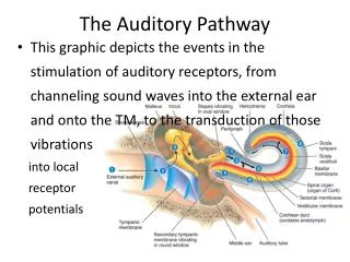
The Ubiquitin Proteosome Pathway. Swati Pradhan Mayura Dange Vidyadhar Daithankar. Overview. Background Protein misfolding & degradation Ubiquitin & proteosome structure Ubiquitin Proteosome Pathway Mechanism Structures of enzymes involved in pathway

E N D
The Ubiquitin Proteosome Pathway Swati Pradhan Mayura Dange Vidyadhar Daithankar
Overview • Background • Protein misfolding & degradation • Ubiquitin & proteosome structure • Ubiquitin Proteosome Pathway • Mechanism • Structures of enzymes involved in pathway • Pathogenic implication of defective pathway • Biological functions of pathway • Diseases & drug development
Post-Translational Modification • Acetylation • Glycosylation • Phosphorylation • Ubiquitination http://www.ncbi.nlm.nih.gov/entrez/query.fcgi?cmdbooks&doptcmdl/Figure+6-79
Degradation of Misfolded Proteins • Lysosomal (extracellular) protein degradation • Protein degraded by lysosomal enzymes • Cytosolic (intracellular) protein degradation • The Ubiquitin Proteosome pathway
Lysosomal degradation • Proteins delivered via endocytosis • Lysosomes • The cellular dust-bins • Contain many hydrolytic enzymes • Proteases • Lipases • Glycosidases
Cytosolic protein degradation • The Ubiquitin Proteosome Pathway www.ihf.de/forschung/ popup/ubiquitin.html
2004 Nobel Prize in Chemistry • The discovery of ubiquitin-mediated protein degradation • Aaron Ciechanover • Avram Hershko • Irwin Rose • Cells give a chemical "kiss of death" to proteins that need to be destroyed.
Targeting by Ubiquitin • Despite help from chaperones, more than 80% fold incorrectly • Proteins are dislocated back into the cytosol • Oligosaccharides are removed • Deglycosylation is catalyzed by N-glycanase • One third of the newly made polypeptide chains are selected for degradation
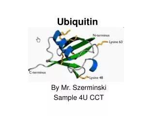
Ubiquitin • 76 amino acids, 8.5 kDa protein • Heat stable • Folds into a compact globular structure • Found throughout the cell • Found in all eukaryotic cells • Human and yeast ubiquitin share 96% sequence identity • Involved in many cellular processes http://www.sanger.ac.uk/Users/sgj/thesis/html/node93.html
The Proteosome • Professional protein degrading organelles • An abundant ATP-dependent protease • Constitutes nearly 1% of cellular protein • Present in many copies throughout the cytosol and the nucleus • Consists of a central hollow cylinder (20S) • Ends of the cylinder are associated with the 19S cap http://walz.med.harvard.edu/Proteasome_Complexes/
The Structure of 20S Proteasome http://www.ncbi.nlm.nih.gov/books/bv.fcgi?rid=stryer.figgrp.3206
Types of Ubiquitination • Mono-ubiquitination • Transcription, histone function, endocytosis and membrane trafficking • Lys48, Lys11 or Lys29 linked poly ubiquitination • Target proteins to the proteasome • Lys63 linked poly ubiquitination • Signaling, DNA repair, stress response, endocytosis and signal transduction
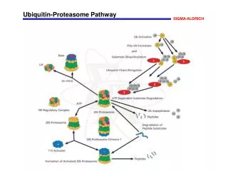
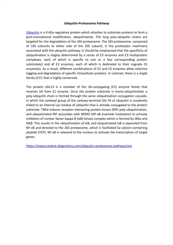
UBIQUITIN PATHWAY • Covalent Attachment of multiple ubiquitin molecules • Degradation of the tagged protein • 3 Enzymes : Ub – Activating enzyme E1 Ub – Conjugating enzyme E2 Ub – Ligases E3
Hierarchical structure • Several E2 transfer Ub from E1 to E3 to which substrate protein is bound • E3s catalyze covalent attachment to the substrate and recognize the substrate
Ubiquitin Activating Enzyme E1 • Adenylation • Thio-ester bond formation • E2 association
Mechanism • E1 activates C-terminus of Ub by forming acyl -adenylate intermediate • Catalytic Cys residue forms thioester bond with Ub • Another Ub is adenylated • Transfer of Ub to E2 forming a thioester bond
Ubiquitin Conjugating enzyme E2 • Carries activated Ub from E1 to the substrate • Cys residue positioned in a shallow groove • Relatively inflexible structure • Conserved Asn may be required for H- bond network OR plays a catalytic role in isopeptide bond formation
Ub Ligases E3 • Final target selection and specificity • Place activated Ub near Lys of substrate • Isopeptide formation of Gly of Ub with the є –NH2 Lys or to the N-terminal residue of the substrate
Categories of E3 Ligases • HECT domain: Homologous to E6-AP C terminus • RING domain: Really Interesting New Gene
HECT Ub Ligases E3 • Conserved 350 amino acids • Catalytic contribution • Forms thiol ester bond with Ub before transferring it to the substrate • N lobe and C- lobe form ‘L’ or ‘inverted T’ shape • Flexibility of hinge loop is required for catalytic activity • C lobe accepts Ub form E2 and transfers it to the substrate • Sequential addition / Indexation
RING Ub Ligases E3 • 15th most common domain in Human genome • Conserved Cys and His Zn2+ co-ordinating residues • Interact directly with E2s • Allosterically activate E2 enzymes • Acts as molecular scaffold • Brings Ub-E2 and substrate closer • Increase # Lys in the vicinity of E2
Polyubiquitination • Poly Ub chain synthesized by adding Ub moieties to Lys of the previous Ub • Another enzyme E4 may be catalyzing this step
Deubiquitination • Thiol proteases • Ubiquitin processing (UBP) enzymes • Removes Ub from polyubiquinated proteins • Ubiquitin carboxy terminal hydrolases (UBH) • Regenerates monomeric Ub
Pathological implication of defective ubiquitin-proteosome pathway
Ubiquitin proteasome pathway is ubiquitous & targets many processes and substrates. • Several complex processes are mediated via degradation or processing of specific proteins. • Aberrations in these systems associates with pathogenic conditions either directly or indirectly.
Pathological Conditions Associated withUbiquitin Proteosome Pathway • Malignancies • Neurodegenerative disorders • Genetic disease Cystic fibrosis, Angelman’s syndrome & Liddle’s syndrome • Immune and inflammatory responses
Malignancies • Oncoproteins like NMyc, c-Myc, c-Fos, are substrates of U-P pathway. • Destabilization of tumor suppressor genes like p53 and p27. • Extremely low levels of p53 in uterine cervical carcinoma. • Prostate, Colorectal and breast cancer: • Tumor suppressor protein p27 is CDK inhibitor of the cell cycle. • Healthy individuals have high levels of p27. Mitogenic stimuli rapidly degrades the protein. • Cancer patients has low levels of p27 in quiescent cells. • Defects in ubiquitin system accelerates degradation of suppressor. • Strong correlation of low levels of p27 and aggressiveness of cancer.
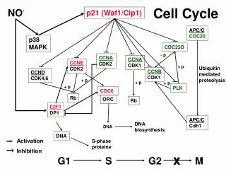
Polyubiquitination Skp2 Defect in ubiquitin pathway ( Skp2) Cell Cycle Regulators and Cancer Degradation of P27
Neurodegenerative disorders • Alzheimer's disease • Parkinson's disease • Huntington’s disease • Spinocerebellar ataxias • Spinobulbar muscular dystrophy (Kennedy’s syndrome) Formation of inclusion bodies (Ref: http://w3.dbb.su.se/~oliveberg/images/bildstrat1.jpg)
Parkinson’s disease and Lewy Bodies ( Ref: http://www.neurodegeneration.uni-goettingen.de/index.html?/en/p311.html) • Accumulation of ubiquitin may be secondary reflecting unsuccessful attempts of ubiquitination. • Abnormal protein associate with each other forming aggregates. • Hypothesis: Aggregated proteins inhibit ubiquitin proteosome pathway.
Liddle’s Syndrome • Hereditary form of hypertension. • Caused due to deletion of proline rich (PY) region in the β and γ subunits of epithelial Na+ channel (hENaC). • HECT domain of E3 binds to PY motif of hENaC. • Mutation in PY motif leads to stabilization of channel complex and E3 ligase • cannot bind to PY motif. • Increased expression of hENaC channel causing excessive reabsorption of sodium and water. Stabilization of channel
Angleman syndrome • Ubiquitin system is considered to be involved in brain development. • Defective synthesis of gene coding for E3 ligase E6-AP • Characteristic symptoms involve mental retardation, seizures, out of context frequent smiling and laughter. • Brain proteins that could be stabilized by mutation have not been identified. Cystic fibrosis • Gene codes for a protein, CFTR, which is chloride ion channel. • Small fraction of protein matures to the cell surface. • Mutation in protein ΔF508, CFTRΔF508 doesn't reach the cell surface. • Ubiquitination degrades mutant CFTRΔF508, resulting in complete lack of cell surface expression.
Ubiquitin proteosome pathway Native protein Foreign protein CLASS I MHC molecule No immune response Immune response • Immune and inflammatory responses • Ubiqutin proteosome pathway is involved in processing of antigenic proteins. • Epitopes are presented on class I MHC molecule generating T cell immune response.
Drug Development for Ubiquitin Dysfunction • Inhibition of enzymes common to entire pathway would target the process non- specifically. • Narrow window between benefits and toxicity needs to be identified. • Develop completely specific E3 ligase inhibitors that would affect the pathways of interests. • Better approach would be development of small molecules that would be specific for substrates.
Conclusions • Ubiquitylation plays a fundamental role of protein degradation at cellular level. (Levels of proteins in nucleus, cytoplasm, ER lumen and transmembrane protein are kept in check by ubiquitin proteosome pathway.) • Ubiquitylation is highly complex, temporally controlled and tightly regulated process. • Enzymologically Ubiquitination is more complex pathway compared to other post translational modification. • Mechanism of catalysis by E3 ligase still remains unclear. • Elucidation of complete catalytic mechanism of ubiquitylation will provide considerable insight on cellular functions.
Extra www.mekentosj.com/ubiquitin/proteasome.html