Download
1 / 13
160 likes | 510 Views
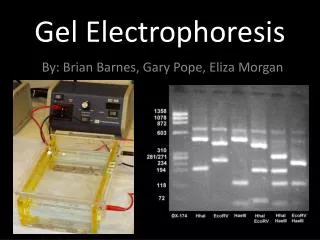
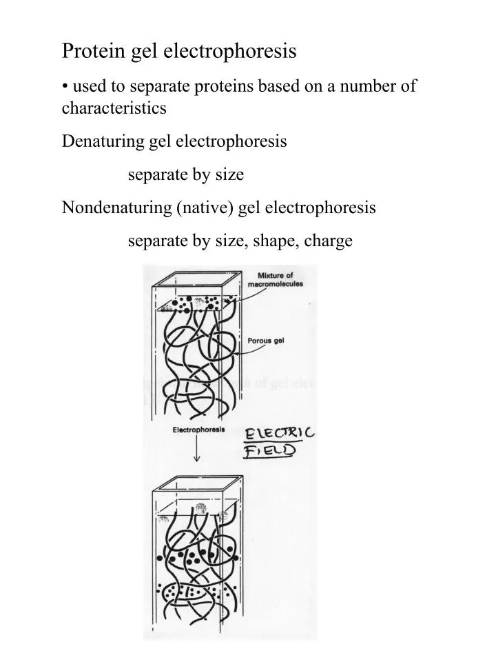
Protein gel electrophoresis • used to separate proteins based on a number of characteristics Denaturing gel electrophoresis separate by size Nondenaturing (native) gel electrophoresis separate by size, shape, charge. Protein gel electrophoresis Polyacrylamide gel electrophoresis.

E N D
Protein gel electrophoresis • used to separate proteins based on a number of characteristics Denaturing gel electrophoresis separate by size Nondenaturing (native) gel electrophoresis separate by size, shape, charge
Protein gel electrophoresis Polyacrylamide gel electrophoresis TEMED - catalyzes free radical formation APS - free radical donor Bisacrylamide - crosslinking agent (19:1 ratio of acrylamide to bis maximizes crosslinking) Higher % of gel - smaller pores (holes) so smaller fragments can be resolved
Protein gel electrophoresis Gradient gels - As proteins migrate through increasing acrylamide concentration, smaller pores, mobility decreases Once proteins reach their “pore limit” little movement and can calc MW
Protein gel electrophoresis Agarose gel electrophoresis Agarose is a long sugar molecule Heated, >70˚ C Room temp Agarose - separation of large molecules 8 kD to 800,000 kD Polyacrylamide - separation of smaller molecules 0.2 kD to 500 kD
Gel electrophoresis Room temp
Protein gel electrophoresis SDS Gel Electrophoresis (agarose or polyacrylamide) Denaturing condition Denature protein by adding SDS (then separate by size only) SDS forms micelles and binds to proteins
Protein gel electrophoresis SDS Gel Electrophoresis (agarose or polyacrylamide) Used to estimate purity and molecular weight, separate proteins by size Electrophoresis of SDS-solvated protein on polyacrylamide gel Stain gel with Coomassie Blue (binds to proteins)
Protein gel electrophoresis Native gel electrophoresis • polypeptides retain their higher-order structure and often retain enzymatic activity and interaction with other polypeptides • migration of proteins depends on many factors, including size, shape, and native charge. • native gels omit the SDS and reducing agent (DTT) • do not put SDS or DTT in the sample buffer • do not heat the samples • prepare the gel and tank buffer solutions without SDS.
Protein gel electrophoresis Separation of hemoglobin proteins Hemoglobin - involved in oxygen transport in body ---------------------------------------------------------- Normal adult Hb (Hb A) - two subunits and 2 subunits Sickle trait hemoglobin (Hb AS) - only one inherited mutation, 50% Hb S and 50% Hb A Sickle hemoglobin (Hb S) - Glu -> Val mutation, Hb S inherited from both parents When Hb S is deoxygenated it crystallizes in RBCs which leads to distortion of red cells and reduction in number of RBCs
Hemoglobin Sickle Cell Anemia Genetic disease in which person inherits gene for sickle-cell Hb from both parents Hb V H L T P E E K Hb-sickle V H L T P V E K Hb-sickle is deoxygenated, insoluble and forms polymers that aggregate Valine has hydrophobic side chain, glutamate has negative charge Valine creates sticky hydrophobic contact point where deoxy-Hb-sickle molecules associate forming long, fibrous aggregates Symptoms: weak, dizzy, short of breath, heart murmurs sickle cells fragile - anemia capillaries blocked -abnormal organ function Patients with sickle cell anemia have to have inherited 2 copies of mutant gene Inherit only 1 copy - resistance to malaria Sickle cell trait ~10% of African American population
Hemoglobin Abnormal Hbs detected in lab by electrophoresis Hb at pH 9.2 has a net (-)charge so moves in electric field toward (+) electrode pI of normal Hb is 6.9 but changes with mutations In Hb S - Glu -> Val ( chain), so 2 fewer (-) charges Example of variations in migration on a gel when Hb mutations present
Sickle Cell Anemia Examine electrophoretic behavior of Hb A, Hb S, Hb AS 1. Set up protein gel (gels are premade) 2. Load Hb samples 3. Separate Hb proteins by applying electric field 4. Take gel apparatus apart 5. Stain gel in Coomassie Blue (entire gel is blue at first) 6. Destain gel (removes nonspecific staining to reveal protein bands on gel)