
Case Presentation
Case presentation of a 13-year-old male with anterior chest wall Ewing's sarcoma, including history, physical examination findings, initial lab investigations, CXR and surgical pathology report.

Case Presentation
E N D
Presentation Transcript
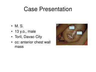
M. S. 13 y.o., male Toril, Davao City cc: anterior chest wall mass Case Presentation
9 mos hx of L anterior chest pain 5 mos persistent pain; palpable mass L ant. chest (~2cms) 1 mo rapid increase in size of mass which later ulcerated associated with purulent foul smelling discharge; consult @ DMC Surgery OPD; excision biopsy was done; result reported as Ewing’s sarcoma Few hours prior to admission, bleeding from mass; admitted at Surgical ward HPI:
Physical examination • Vital signs BP=90/60 RR=21 BEE=1200 kcal/day T=35.5 CR=122 BMI=13.8 kg/m2 • Conscious; coherent; NICRD; afebrile • Anicteric sclerae; pale palpebral conjunctivae; (-) lympadenopathies • Asymmetrical chest expansion; clear breath sounds • (+) 10 x 10 cm nodular, ulcerating mass with purulent foul smelling discharge @ left anterolateral chest • Adynamic precordium; NRRR; (-) murmurs • Flat, soft abdomen; (-) tenderness; (-) palpable masses • Full equal pulses; pale nail beds
CBC WBC 7.73 Hb 160 Hct 0.50 Rbc 5.67 Diff count Neut 80% Lym 13% Mono 7% Protime PT pt 12.4 PT INR 1.03PT % activity 92% PT control 13.3 Initial Laboratory Investigations
CXR result: Anterior chest wall mass, left, with pleural, pulmonary, osseous, axillary lymph nodes metastasis.
Surgical Pathology Report Specimen 6th rib mass Gross description specimen consists of 2.0 cm aggregate of creamy white to brown tissue fragments Microscopic description sections show sheets of small atypical cells showing large hyperchromatic nuclei and very scant cytoplasm. These cells are thrown into poorly formed lobules in some areas by very thin fibrous stroma. Large areas of necrosis are seen among the atypical cells Diagnosis: consistent with Ewing’s sarcoma
Impression:Ewing’s sarcoma stage IV ( T2b, N1,M1 ) anterolateral chest


