Download
1 / 29
300 likes | 429 Views
Comprehensive guidelines for diagnosing and managing DORV, covering anatomy, classifications, diagnostic methods, surgical considerations, and treatment strategies.

E N D
DGPK guidelineDouble Outlet Right Ventricle(DORV) H. Bertram, MHH, HannoverJ. Weil, UKE, HamburgJ. Sachweh, UKE, HamburgDGPK guideline committee
Guideline DORV Definition 100 % + 100 % 100 % + > 50 % Double Outlet Right Ventricle (DORV) represents a spectrum of congenitally malformed hearts in which the circumference of both arterial valves, or the greater part of both circumferences, are supported by the right ventricle prevalence: • 1,3 % of cardiac defects • 1,1 / 10.000 live births (PAN)
Guideline DORV Definition > 150 % rule Mahle WT et al. Cardiol Young 2008; 18(Suppl. 3): 39–51
Guideline DORV Definition Double Outlet Right Ventricle (DORV) represents a spectrum of congenitally malformed hearts in which the circumference of both arterial valves, or the greater part of both circumferences, are supported by the right ventricle • same ventriculo-arterial connection, but variations in - infundibular morphology - arterial interrelationship - coronary arterial anatomy • any arrangement of the atrial appendages, or situs • any atrioventricular connection • multiple combinations of associated malformations
Guideline DORV spatial relationship of the semilunar cusps in hearts with DORV
Guideline DORV ‚Interventricular communication‘ vs ‚VSD‘ Mahle WT et al. Cardiol Young 2008; 18(Suppl. 3): 39–51
Guideline DORVDiagnostics • Goal: displaying cardiac anatomy with emphasis on potential surgical biventricular repair (feasibility of tunneling the interventricular communication to one or other arterial trunk) • position, size, interrelationship, course of the great arteries • morphology and size of the interventicular communication / the VSD in relation to diameter of the aortic valve • location and severity of a subpulmonary or subaortic obstruction • morphology and size of both ventricles and AV-valves • Methods: • Echocardiography• Angiography (Cath./MRT/CT)
Guideline DORV • Classification of the interventricular communication / VSD according to its location in relation to the great arteries • subaortic (65%) • subpulmonary (20-25%) • doubly committed (3%) • non committed (7%)
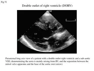
Guideline DORVsubaortic VSD • subaortic VSD • size in relation to the diameter of the aorta • distance between VSD and aortic valve • presence and severity of a subpulmonary obstruction • subcostal coronal and sagittal planes; parasternal long axis
Guideline DORVsubaortic VSD parasternal long axis Ao Ao LV LA
Guideline DORVsubaortic VSD Subcostal TEE RV Ao PA
Guideline DORVsubaortic VSD subaortic VSD with severe subpulmonary obstruction RV RV Ao PA Ao
Guideline DORV • doubly committed VSD • size and distance of the VSD to the aorta / pulmonary artery • presence and severity of a subpulmonary obstruction • subcostal coronal and sagittal planes
Guideline DORV • subpulmonary VSD • size in relation to the diameter of the pulmonary artery • presence and severity of a subpulmonary obstruction • subcostal coronal and parasternal long axis planes
Guideline DORVsubpulmonary VSD RV PA RV PA
Guideline DORVsubpulmonary VSD • Taussig-Bing malformation: • DORV with subpulmonary VSD • semilunar valves side-by-side • no subpulmonary obstruction • semilunar valves and AV-valves separated by conal septum
Guideline DORV • non-committed VSD • location and size; distance to semilunar valves • presence and severity of a subpulmonary obstruction • subcostal coronal / 4 C views LV Ao
Guideline DORVnon-committed VSD RV PA LV Ao RV
Guideline DORV Double Outlet Right Ventricle Malposition of the great arteries, which arise completely (100% + 100%) or with the greater part of their circumference (100% + > 50%) from the right ventricle interventricular communication in relation to the great arteries ‚doubly committed‘ subpulmonary VSD ‚non committed‘ AVSD / heterotaxy subaortic VSD • right atrial isomerism • TAPVD • l-SVC • subpulmonary obstruction concommittant malformations aortic coarctation (in ~ 50 %) subpulmonary obstruction valvular / subvalvular PS valvular / subvalvular PS clinical symptoms pulmonary hyperperfusion;congestive heart failure reduced lung perfusion;mild severe cyanosis severe cyanosis; congestive heart failure clinical symptoms determined by concommittant malformations clinical subtype TOF - type VSD - type TGA - type complex DORV biventricular repair 2-6 years(complex intracardiac tunneling +/- VSD incision / arterial switch) ordefinitive univentricular palliation biventricular repair < 1 mo(VSD-closure + arterial switch) biventricular repair 4-12 mo(VSD closure + relief of RVOTO) biventricular repair 1-6 mo(VSDclosure) surgical strategy
Guideline DORV • medical treatment • PG E in duct dependent pts with severe subpulmonary obstruction • diuretics, ß-blockers, … in pts with pulmonary hypercirculation and congestive heart failure • catheter intervention • TOF-type: balloon valvuloplasty; ductal stent; RVOT stent • TGA-type: BAS • surgical palliation • TOF-type: modified BT-shunt if primary repair is not suitable or considered ‚high risk‘ • PAB in ncVSD to delay complex intraventricular repair
Guideline DORV Surgical Repair VSD - type • VSD closure in the age of 1 to 6 months- some pts need enlargement of the VSD (> 4/5 aortic annulus); cave: AV-Block TOF - type • VSD closure and relief of subpulmonary obstruction in the age of 4 to 12 months+/- muscular and transjunctional incision or patch enlargement
Guideline DORV Surgical Repair TGA - type • neonatal corrective surgery with arterial switch, VSD closure+/- resection of aortic coarctation / aortic arch reconstruction +/- resection of subaortic infundibulum cave: coronary artery anomalies • alternatively ‚Rastelli - type repair‘: baffling of the left ventricle to both arterial valves and placement of a conduit from RV to the pulmonary trunk Subpulmonary VSD with valvular/subvalvular pulm. stenosis • ‚Kawashima-OP‘ • ‚Rastelli-OP‘ • ‚REV-procedure‘ (Reparation a l‘ Etage Ventriculaire) • ‚Aortic translocation‘ – ‚Nikaidoh-procedure‘
Guideline DORV Surgical Repair Complex DORV • biventricular repair aged 2-6 years: complex intraventricular baffling (LV Ao/PA) +/- VSD enlargement +/- arterial switch • definitive functionally univentricular palliation
Guideline DORV Surgical Repair Implications of the 200 % rule (‚true‘ DORV) Percent of Great Vessels Arising from the RV Modified from: F. Lacour-Gayet: Intracardiac Repair of Double Outlet Right Ventricle Semin Thorac Cardiovasc Surg Pediatr Card Surg Ann 2008;11:39-43
Guideline DORV prognosis • biventricular repair achievable in most pts • increased operative risk determined by concommittant malformations: • aortic arch obstructions • AV-valve anomalies• coronary arterial anomalies • LV hypoplasia • multiple VSDs
Guideline DORV Residual lesions requiring reinterventions after surgical repair • Depending on different morphology and type of previous surgical repair • TOF – type: - pulmonary valve incompetence - residual subpulmonary obstruction • RV-PA-conduit: definitive reoperation for conduit replacement(stenosis, incompetence, size-mismatch in growing children) • complex intracardiac baffling: subaortic obstruction biventrcular surgical repair has a much higher rate of reintervention than a strategy of functionally univentricular palliation
Guideline DORV Follow-up • Life-long follow-up by pediatric cardiologists and subsequently specialists for adult congenital heart disease is mandatory