Neuroglia
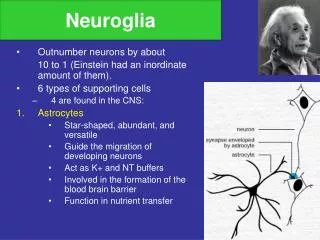
Neuroglia. Outnumber neurons by about 10 to 1 (Einstein had an inordinate amount of them). 6 types of supporting cells 4 are found in the CNS: Astrocytes Star-shaped, abundant, and versatile Guide the migration of developing neurons Act as K+ and NT buffers

Neuroglia
E N D
Presentation Transcript
Neuroglia • Outnumber neurons by about 10 to 1 (Einstein had an inordinate amount of them). • 6 types of supporting cells • 4 are found in the CNS: • Astrocytes • Star-shaped, abundant, and versatile • Guide the migration of developing neurons • Act as K+ and NT buffers • Involved in the formation of the blood brain barrier • Function in nutrient transfer
Neuroglia • Microglia • Specialized immune cells that act as the macrophages of the CNS • Why is it important for the CNS to have its own army of immune cells? • Ependymal Cells • Low columnar epithelial-esque cells that line the ventricles of the brain and the central canal of the spinal cord • Some are ciliated which facilitates the movement of cerebrospinal fluid
Neuroglia 4. Oligodendrocytes • Produce the myelin sheath which provides the electrical insulation for certain neurons in the CNS
Neuroglia • 2 types of glia in the PNS • Satellite cells • Surround clusters of neuronal cell bodies in the PNS • Unknown function • Schwann cells • Form myelin sheaths around the larger nerve fibers in the PNS. • Vital to neuronal regeneration
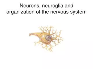
Neurons • The functional and structural unit of the nervous system • Specialized to conduct information from one part of the body to another • There are many, many different types of neurons but most have certain structural and functional characteristics in common: • Cell body (soma) • One or more specialized, slender processes (axons/dendrites) • An input region (dendrites/soma) • A conducting component (axon) • A secretory (output) region (axon terminal)
Soma • Contains nucleus plus most normal organelles. • Biosynthetic center of the neuron. • Contains a very active and developed rough endoplasmic reticulum. • The neuronal rough ER is referred to as the Nissl body. • Contains many bundles of protein filaments (neurofibrils) which help maintain the shape, structure, and integrity of the cell. In the soma above, notice the small black circle. It is the nucleolus, the site of ribosome synthesis. The light circular area around it is the nucleus. The mottled dark areas found throughout the cytoplasm are the Nissl substance.
Somata • Contain multiple mitochondria. • Acts as a receptive service for interaction with other neurons. • Most somata are found in the bony environs of the CNS. • Clusters of somata in the CNS are known as nuclei. Clusters of somata in the PNS are known as ganglia.
Neuronal Processes • Armlike extensions emanating from every neuron. • The CNS consists of both somata and processes whereas the bulk of the PNS consists of processes. • Tracts = Bundles of processes in the CNS (red arrow) Nerves = Bundles of processes in the PNS • 2 types of processes that differ in structure and function: • Dendrites and Axons
Dendrites are thin, branched processes whose main function is to receive incoming signals. • They effectively increase the surface area of a neuron to increase its ability to communicate with other neurons. • Small, mushroom-shaped dendritic spines further increase the SA • Convey info towards the soma thru the use of graded potentials – which are somewhat similar to action potentials. Notice the multiple processes extending from the neuron on the right. Also notice the multiple dark circular dots in the slide. They’re not neurons, so they must be…
Most neurons have a single axon – a long (up to 1m) process designed to convey info away from the cell body. • Originates from a special region of the cell body called the axon hillock. • Transmit APs from the soma toward the end of the axon where they cause NT release. • Often branch sparsely, forming collaterals. • Each collateral may split into telodendria which end in a synaptic knob, which contains synaptic vesicles – membranous bags of NTs.
Axons • Axolemma = axon plasma membrane. • Surrounded by a myelin sheath, a wrapping of lipid which: • Protects the axon and electrically isolates it • Increases the rate of AP transmission • The myelin sheath is made by Oligodendrocytes in the CNS and by Schwann cells in the PNS. • This wrapping is never complete. Interspersed along the axon are gaps where there is no myelin – these are nodes of Ranvier. • In the PNS, the exterior of the Schwann cell surrounding an axon is the neurilemma
Myelination in the CNS Myelination in the PNS
A bundle of processes in the PNS is a nerve. • Within a nerve, each axon is surrounded by an endoneurium(too small to see on the photomicrograph) – a layer of loose CT. • Groups of fibers are bound together into bundles (fascicles) by a perineurium (red arrow). • All the fascicles of a nerve are enclosed by a epineurium (black arrow).
Types of Nerve Fibers • Group A • Axons of the somatic sensory neurons and motor neurons serving the skin, skeletal muscles, and joints. • Large diameters and thick myelin sheaths. • Group B • Type B are lightly myelinated and of intermediate diameter. • Group C • Type C are unmyelinated and have the smallest diameter. • Autonomic nervous system fibers serving the visceral organs, visceral sensory fibers, and small somatic sensory fibers are Type B and Type C fibers.
What is Blood Brain Barrier? • The BBB is formed by the single layer of endothelial cells that line the inner surfaces of capillaries in the brain. • It is a semi-permeable capillary membrane; that is, it allows some materials to cross, but prevents others from crossing. In most parts of the body the capillaries, are lined with endothelial cells. The endothelial tissue has small spaces between each individual cell so substances can move readily between the inside and the outside of the vessel. However, in the brain, the endothelial cells fit tightly together and substances cannot pass out of the bloodstream. (Some molecules, such as glucose, are transported out of the blood by special methods such as active transport.)
What is the Blood Brain Barrier? • Structural and functional barrier which impedes and regulates the influx of most compounds from blood to brain • Formed by brain microvascular endothelial cells (BMEC), astrocyte end feet and pericytes • Essential for normal function of CNS • Regulates passage of molecules in and out of brain to maintain neural environment. • Responsible for metabolic activities such as the metabolism of L-dopa to regulate its concentration in the brain.
Structure of Blood Brain Barrier Source: Bock et al
Differences between BMEC and normal endothelial cells • Structural differences: • Absence of fenestrations • More extensive tight junctions (TJ) • Functional differences: • Impermeable to most substances • Sparse pinocytic vesicular transport • Increased expression of transport and carrier proteins: receptor mediated endocytosis • No gap junctions, only tight junctions • Limited paracellular and transcellular transport
Functions and Properties of the BBB • The BBB has several important functions: • Protects the brain from "foreign substances" in the blood that may injure the brain. • Protects the brain from hormones and neurotransmitters in the rest of the body. • Maintains a constant environment for the brain.
Functions and Properties of the BBB • General Properties of the BBB • Large molecules do not pass through the BBB easily. • Low lipid (fat) soluble molecules do not penetrate into the brain. However, lipid soluble molecules rapidly cross the BBB into the brain. • Molecules that have a high electrical charge to them are slowed. • Therefore: • The BBB is selectively permeable to :Oxygen, Carbon dioxide and glucose • The BBB is not permeable to hydrogen ions
Integrity of BBB • Tight Junctions • Adherens Junctions • Pericytes • Astrocyte end feet
Tight Junctions between BMEC • Appear at sites of apparent fusion between outer leaflets of plasma membrane of endothelial cells • Continuous • Anastomosing • Intramenbranous strands or fibrils on P face with complementary groove on E face • Protein components: • Claudin • Occludin • Junction Adhesion Molecules • Accessory proteins Source: Ballabh et al
Claudin • 22kDa phosphoprotein • 4 transmembrane domains • localized in TJ strands Source: Ballabh et al
Occludin • 65kDa phosphoprotein, • 1° structure very different from claudin • Regulatory proteins: alters paracellular permeability. Source: Ballabh et al
Barrier Function of Occludin and Claudin • Assemble into heteropolymers and form intramembranous strands which contain channels allowing selective diffusion of ions and hydrophilic molecules. • Breakdown of BBB in tissue surrounding brain tumors occurs with concomitant loss of 55kDa occludin expression
Junction Adhesion Molecules: • 40kDa • Integral membrane protein, single transmembrane region • Belongs to immunoglobulin superfamily • Localizes at tight junctions • Involved in cell-to-cell adhesion and monocyte transmigration through BBB • Regulates paracellular permeability and leukocyte migration • Also found on circulating leukocytes, platelets and lymphoid organs.
BMEC intercellular space Source: Ballabh et al
Barrier function of JAM • Homotypic binding between JAM molecules on adjacent endothelial cells acts as a barrier for circulating leukocytes • Heterotypic binding of endothelial JAM to leukocyte JAM might guide transmigration of leukocytes across interendothelial junctions • So factors that decrease leukocyte migration must either strengthen homotypic interactions or weaken heterotypic interactions.
Cytoplasmic accessory proteins • (ZO-1, ZO-2, ZO-3, cingulin etc) • These link membrane proteins to actin • maintenance of structural and functional integrity of endothelium • crosslink transmembrane proteins. • Membrane associated guanylate kinase-like proteins (MAGUKS) • subunits function as protein binding molecules • role in organization the plasma membrane
Adherens Junction • Complex between membrane protein cadherin and intermediary proteins called catenins • Cadherin-catenin complex joins to actin cytoskeleton • Form adhesive contacts between cells. • Assemble via homophilic interactions between extracellular domains of calcium ion dependent cadherins on surface of adjacent cells
Pericytes: • Cells of microvessels including capillaries, venules, and arterioles that wrap around endothelial cells. • Provide structural support and vasodynamic capacity to microvasculature. • Role in structural stability of vessel wall • Endothelial cells associated with pericytes are more resistance to apoptosis than isolated endothelial cells • Indicates role of PC in structural integrity and genesis of the BBB • Phagocytic activity
Astrocyte end feet • Star shaped glial cells • Provides biochemical support for BMEC • Influence of morphogenesis and organization of vessel wall • Factors released by astrocytes involved in postnatal maturation of BBB • Direct contact between endothelial cells and astrocytes necessary to generate BBB (Rubin et al, 1991) • Co-regulate function by the secretion of soluble cytokines such as (LIF, leukemia inhibiting factor), Ca2+ dependent signals by intracellular IP-3 and gap junction dependent pathways, and second messenger pathways involving extracellular diffusion of purinergic messenger.
Regions of brain not enclosed by BBB • Circumventricular organs • area postrema, • median eminence, • neurohypophysis, • pineal gland, • subfornical organ and • lamina terminalis These are regions which need to respond to factors present in systemic circulation
Transport at the BBB • There are five basic mechanisms by which solute molecules move across membranes: • simple diffusion • facilitated diffusion • simple diffusion through an aqueous channel • active transport through a protein carrier • Endocytosis
Transport mechanisms at the BBB. 1 = paracellular diffusion , 2 = transcellular diffusion , 3 = ion channel 4 = ion-symport channel 5 = ion-antiport channel 6 = facilitated diffusion , 7 = active efflux pump 8 = active-antiport transport , 9 = receptor mediated endocytosis
Diffusion • Phospholipid bilayer • Movement of substances down diffusion gradient • Transfer of lipophilic substances • alcohol, nicotine, oxygen, carbon dioxide
Facilitated transport • Carrier systems • particular essential amino acids, glucose, these are extremely specific • transport D-glucose only, • large neutral amino acids which act as precursors for neurotransmitters, • only which the brain cannot make, • glycine: it can block the transmission of nerve signals, hence special carrier which ensures that glycine can be removed from brain • Receptor mediated endocytosis • Leptin, insulin, overlaps with carrier systems