Gallstone disease
Gallstone disease. doc. MUDr. Radan Keil, PhD. . Definitions. Cholecystolithiasis – biliary stones in the gallbladder Choledocholithiasis – biliary stones int the common bile duct and hepatic duct. Biliary stones .

Gallstone disease
E N D
Presentation Transcript
Gallstone disease doc. MUDr. Radan Keil, PhD.
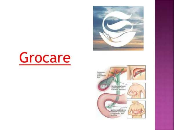
Definitions • Cholecystolithiasis – biliary stones in the gallbladder • Choledocholithiasis – biliary stones int the common bile duct and hepatic duct.
Biliary stones • Cholesterol stones – 90% of all concrements – made from cholesterol and calcium • Black pigment stones made from calciumbilirubinate /hemolytic anemias, liver cirrhosis/ • Brown pigment stones – after infection in biliary tract – made from unconjugated bilirubin cholesterol and proteins
Incidence • 10% of population – in western countries more • Women 2-3x more frequent
Risk factors • Age/cholesterol +, bileacids-/ • Gender /women + /cholesterol +/ • Obesity /cholesterol +/ • Weightloss/cholesterol+,hypomotilityofgallbladder/ • Pregnancy/dtto/ • Drugs • Geneticpredispozition/cholesterol +/ • Ilnessofterminal ileum, hyperlipoproteinemiasIIb,IV • Hemolyticanemia • G negative infection in thebileduct
Clinical presentation • Biliary colic is a common manifestation of gallstone disease. • Patients suffer from intermitent colic pain in the right upper abdominal quadrant. • Biliary colic occurs due to intermitent obstruction of the cystic duct by stones.
Physical examination • Positive Murphys sign • Mild or moderate right upper quadrant tenderness .
Methods of investigation • Laboratory findings - liver tests mostly normal. • Blood count – mostly normal, light leucocytosis is possible.
Methods of investigation • Abdominal ultrasound is the first method of choice. • Should be performed to asses the gallstones in the gallbladder or in another part of biliary tract.
Diferential diagnosis • Renal colic • Acute pancreatitis • Peptic ulcer disease • Ischemic heart disease • Right sided pneumonia
Therapy • It is necessary to divide patienst into three groups • 1/asymptomatic patients -60-80% • 2/symptomatic patients without complications • 3/symptomatic patients with complications
Asymptomatic patients • You should recommend low fat intake, diet regimen. • It is necessary to inform the patient, that in case of biliary colic he should come.
Symptomatic patient without complication • This patient should undergo cholecystectomy. • In this case it is prevention of further complications /30% in next 20 years/
Symptomatic patients with complication /cholangitis, cholecystitis/ • For the first it is necessary to solve the problem . • In the case of cholangitis patients should undergo acute ERCP with extraction of the stones and then as soon as possible it is necessary to provide cholecystectomy
Cholecystitis • Admition to the hospital • Nothing orally • Antibiotics • Intravenous hydratation • Analgetics
Cholecystitis • After six weeks patients should undergo cholecystectomy.