Download
1 / 28
280 likes | 317 Views
Learn about the resting potential, generation, and conduction of action potentials in neurons. Explore how signal transmission occurs and the importance of synapses in neural communication.

E N D
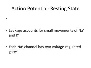
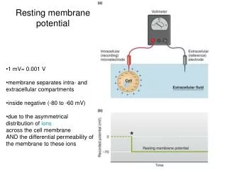
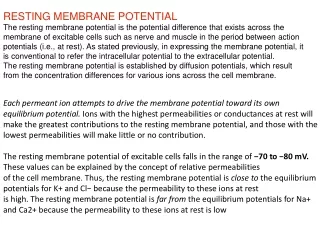
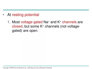
At resting potential • Most voltage-gated Na+ and K+channels are closed, but some K+ channels (not voltage-gated) are open.
When an action potential is generated • Voltage-gated Na+ channels open first and Na+ flows into the cell. • During the rising phase, the threshold is crossed, and the membrane potential increases. • During the falling phase, voltage-gated Na+ channels become inactivated; voltage-gated K+ channels open, and K+ flows out of the cell. • Cell is now repolarizedbut is not normal until Na+ K+ pumprestores original resting potential.
During the refractory period after an action potential, a second action potential cannot be initiated. This ensures that an impulse moves along the axon in one direction only. • The refractory period is a result of a temporary inactivation of the Na+ channels. • The refractory period is a period of “normal” repolarization when the Na+ K+ pumprestores the membrane to its original polarized condition.
Conduction of Action Potentials • An action potential can travel long distances by regenerating itself along the axon. • At the site where the action potential is generated, usually the axon hillock, an electrical current depolarizes the neighboring region of the axon membrane. • Inactivated Na+ channels behind the zone of depolarization prevent the action potential from traveling backwards. Action potentials travel in only one direction: toward the synaptic terminals.
Conduction of an Action Potential Signal Transmission Axon Plasma membrane Action potential Cytosol Na+ Action potential K+ Na+ K+ Action potential K+ Na+ K+
Conduction Speed • The speed of an action potential increases with the axon’s diameter. • In vertebrates, axons are insulated by a myelin sheath, which causes an action potential’s speed to increase. • Myelin sheaths are made by glia— oligodendrocytes in the CNS and Schwann cellsin the PNS.
Schwann cells and the myelin sheath Node of Ranvier Layers of myelin Axon Schwann cell Schwann cell Nodes of Ranvier Nucleus of Schwann cell Myelin sheath Axon
Action potentials are formed only at nodes of Ranvier, gaps in the myelin sheath where voltage-gated Na+ channels are found. • Action potentials in myelinated axons jump between the nodes of Ranvier in a process called saltatory conduction.
Saltatory conduction Schwann cell Depolarized region (node of Ranvier) Cell body Myelin sheath Axon
Neurons communicate with other cells at synapses • At electrical synapses, the electrical current flows from one neuron to another. • At chemical synapses, a chemical neurotransmitter carries information across the gap junction = synapse. • Most synapses are chemical synapses.
The presynaptic neuron synthesizes and packages the neurotransmitter in synaptic vesicleslocated in the synaptic terminal. • The action potential causes the release of the neurotransmitter. • The neurotransmitter diffuses across the synaptic cleft and is received by the postsynaptic cell.
Chemical synapse 5 Na+ K+ Synaptic vesicles containing neurotransmitter Presynaptic membrane Voltage-gated Ca2+ channel Postsynaptic membrane Ca2+ 1 4 6 2 3 Synaptic cleft Ligand-gated ion channels
Generation of Postsynaptic Potentials • Direct synaptic transmission involves binding of neurotransmitters to ligand-gated ion channelsin the postsynaptic cell. • Neurotransmitter binding causes ion channels to open, generating a postsynaptic potential.
Postsynaptic potentials fall into two categories: • Excitatory postsynaptic potentials (EPSPs) are depolarizations that bring the membrane potential toward threshold. • Inhibitory postsynaptic potentials (IPSPs) are hyperpolarizations that move the membrane potential farther from threshold.
After release, the neurotransmitter • May diffuse out of the synaptic cleft • May be taken up by surrounding cells • May be degraded by enzymes
Summation of Postsynaptic Potentials • Unlike action potentials, postsynaptic potentials are graded and do not regenerate. • Most neurons have many synapses on their dendrites and cell body. • A single EPSP is usually too small to trigger an action potential in a postsynaptic neuron. • If two EPSPs are produced in rapid succession, an effect called temporal summation occurs.
Summation of postsynaptic potentials Terminal branch of presynaptic neuron E1 E1 E1 E1 E2 E2 E2 E2 Axon hillock Postsynaptic neuron I I I I 0 Action potential Action potential Threshold of axon of postsynaptic neuron Membrane potential (mV) Resting potential –70 E1 + I I E1 E1 E1 E1 E1 E1 + E2 (b) Temporal summation (d) Spatial summation of EPSP and IPSP (a) Subthreshold, no summation (c) Spatial summation
In spatial summation, EPSPs produced nearly simultaneously by different synapses on the same postsynaptic neuron add together. The combination of EPSPs through spatial and temporal summation can trigger an action potential. • Through summation, an IPSP can counter the effect of an EPSP. Thesummed effect of EPSPs and IPSPs determines whether an axon hillock will reach threshold and generate an action potential.
Modulated / Indirect Synaptic Transmission • In indirect synaptic transmission, a neurotransmitter binds to a receptorthat is not part of an ion channel. • This binding activatesa signal transduction pathwayinvolving a second messenger in the postsynaptic cell. • Effects of indirect synaptic transmission have a slower onset but last longer.
Neurotransmitters • The same neurotransmitter can produce different effects in different types of cells. • There are five major classes of neurotransmitters: acetylcholine, biogenicamines, amino acids, neuropeptides, andgases. • Gases such as nitric oxide and carbon monoxide are local regulators in the PNS.
Acetylcholine • Acetylcholineis a common neurotransmitter in vertebrates and invertebrates. • In vertebrates it is usually an excitatory transmitter. • Common at the neuro-muscular junction.
Biogenic Amines & Amino Acids • Biogenic amines include epinephrine, norepinephrine, dopamine, and serotonin. They are active in the CNS and PNS. • Two amino acids are known to function as major neurotransmittersin the CNS: gamma-aminobutyric acid (GABA) and glutamate.
Neuropeptides • Several neuropeptides, relatively short chains of amino acids, also function as neurotransmitters. • Neuropeptides include substance P and endorphins,which both affect our perception of pain. • Opiates bind to the same receptors as endorphins and can be used as painkillers.
Review Action potential +50 Falling phase 0 Rising phase Membrane potential (mV) Threshold (–55) –50 Resting potential –70 Depolarization Undershoot –100 Time (msec)
You should now be able to: • Distinguish among the following sets of terms: sensory neurons, interneurons, and motor neurons; membrane potential and resting potential; ungated and gated ion channels; electrical synapse and chemical synapse; EPSP and IPSP; summation. • Explain the role of the sodium-potassium pump in maintaining the resting potential.
Describe the stages of an action potential; explain the role of voltage-gated ion channels in this process. • Explain why the action potential cannot travel back toward the cell body. • Describe saltatory conduction. • Describe the events that lead to the release of neurotransmitters into the synaptic cleft.
Explain the statement: “Unlike action potentials, which are all-or-none events, postsynaptic potentials are graded.” • Name and describe five categories of neurotransmitters.