Download
1 / 61
610 likes | 629 Views
Learn about Enteric and Non-Enteric rods, including specific characteristics, cultural appearances, and biochemical tests in the lab setting. Understand the identification and differentiation of Klebsiella, Proteus, Salmonella, Shigella, and their roles in infections. Explore microscopic examinations, colony appearances on different media, and key biochemical tests. Discover the significance of these bacteria in human health and potential diseases they can cause.

E N D
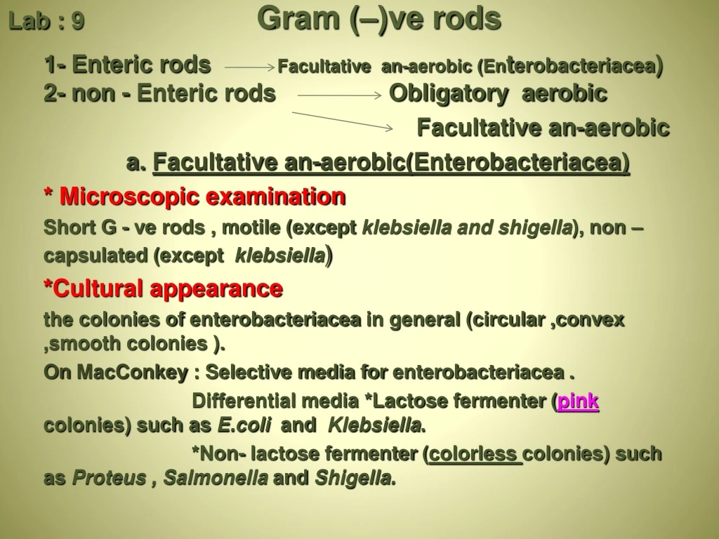
Lab : 9 Gram (–)ve rods 1- Enteric rods Facultative an-aerobic (Enterobacteriacea) 2- non - Enteric rods Obligatory aerobic Facultative an-aerobic a. Facultative an-aerobic(Enterobacteriacea) * Microscopic examination Short G - ve rods , motile (except klebsiella and shigella), non –capsulated (except klebsiella) Cultural appearance * the colonies of enterobacteriacea in general (circular ,convex ,smooth colonies ). On MacConkey: Selective media for enterobacteriacea . Differential media *Lactose fermenter (pinkcolonies) such as E.coli and Klebsiella. *Non- lactose fermenter (colorless colonies) such as Proteus , Salmonella and Shigella.
Escherichia coli It is found as normal flora in large intestine of most humans , though usually benign , some strains cause traveler's diarrhea , even benign strains can cause urinary tract infections Microscopic examination Gram ( - ) ve rods ,straight with rounded ends ,mostly are motile . Cultural appearance * Nutrient agar : Opaque , smooth,convex,with entire edges . Blood agar : some produce β –hemolysis . Eosin methylene blue ( EMB ) : metalic green sheen . Mac Conkyagar : pink (lactose Fe.) , non viscous colonies . Biochemical test ٍ (TSI) test : Acid slant (+)ve* Acid butt (+)ve With gas production , but no H2S produced . IMVC : + + - - Urease : ( - ) ve Nitrate reduction : (+)ve
MacConkey Agar Differential Media: left: no lactose fermentation right: lactose fermentation metalic green on ( EMB )
Klebsiella It is found as normal flora (large intestine , urethra) , human feces, soil, water, grain, fruits, vegetables , may cause pediatric septicemia , pneumonia , urinary tract infections • Microscopic examination Gram ( - ) ve rods , capsulated , non motile . Cultural appearance * Blood agar : gray, large colonies , circular , entire, high convex, often mucoid on primary isolation.. Mac Conky agar: usually pink in center with clear edge (lactose Fer.) Biochemical test (TSI) test : Acid slant (+)ve* Acid butt (+)ve With gas production no H2S produced IMVC : - - + + Urease :( + ) ve Nitrate reduction : (+) ve
Proteus It is found as normal flora (large intestine , urethra), manure; soil; polluted waters, Only cause infection when leave intestinal tract (Opportunist) may cause urinary tract and wound Infection (especially burns) ; infantile diarrhea * Microscopic examination Gram ( - ) ve rods , it is highly motile . Cultural appearance Fishy smell and swarming appearance. Blood agar : an irregular flat colony that can rapidly spread across the surface of a fresh plate . Mac Conky agar: flat irregular colorless colonies, may spread on fresh medium .
Biochemical test (TSI) test : alk slant (-) or Acid slant (+)ve for P vulgaris* Acid butt (+)ve With gas production , H2S produced . IMVC : v +-vUrease : (+) ve Nitrate reduction : (+) ve Phenylalanine deaminase test : (+) ve Proteus vulgaris
Salmonella Transmitted from animal to human by the oral rout ( milk ) ,it result in enteritis systemic infection ,food poisoning ( Salmonella typhimurium) , typhoid fever (Salmonella typhi) , or para typhoid fever (Sal. paratyphi A , Sal. paratyphi B) . * Microscopic examination Gram ( - ) ve rods Cultural appearance* circular ,smooth , entire edge ,. Mac Conky agar: pale colonies (non lactose fer.). ●Bismuth Sulfite agar ( BSA ) : black to brown colonies with metallic sheen appearance , because of H2S production . Salmonella Shigella agar (SSA): colorless colonies with black centers .
Biochemical test (TSI) test : Alk slant (-) ve * Acid butt (+) ve gas or No gas production , H2S produced . IMVC : v+ - - Urease : (-) ve Nitrate reduction : (+) ve Sal.Typhi -/+/+ Sal. Typhimurium
Bismuth sulfate agar is selective for Salmonella spp.
Widal test(indirect latex agglutination test) • Is used to detect antibodies against H and O antigens of the Salmonella typhiand paratyphi. The significant Abs titer for both anti-O and anti-H is 1/160 or more.
Shigella It is normal flora (large intestine),may cause bacillary dysentery (shigellosis ); traveler's diarrhea. S. dysenteriaeis the agent of classic bacterial dysentery * Microscopic examination Gram ( - ) ve rods ,non motile Cultural appearance * smooth ,convex , entire edge , transparent . Mac Conky agar: pale colonies (non lactose fer.). Salmonella Shigella agar (SSA): colorless colonies without black centers .
Biochemical test (TSI) test : Alk slant (-) ve * Acid butt (+) ve No gas production , no H2S produced . IMVC : v + - - Urease : (-) ve Nitrate reduction : (+) ve
G G+ve rods Non sporeforming Tetanus e.g. Cl. tetani Spore forming o2 co2 Bacillus anthracis corynebacterium Diphtheria Bacillus subtilis Bacillus cereus Clostridium perfringens Clostridium tetani Cl. botulinum Cl. difficille
Corynebacteriumdiphtheriae Corynebacterium diphtheriae is a pathogenic bacterium that causes diphtheria. It is also known as the Klebs-Löffler bacillus Microscopic examination * Gram +ve rods * pleomorphic (Chinese letter,V, palisades arrangment) * Non spore forming * non motile * non capsulated *Metachromatic granules (volutin granules) * Blue biplar with albert stain
Macroscopic appearance Cultural appearance Black colony on tellurite blood agar (Tinsdales agar) due to the production of H2S from cystine • biochemical test Gelatin -ve Oxidase +ve Starch +ve Glucose +ve Maltose +ve
Bacillus anthracis Bacillus anthracisis a pathogenic bacterium that causes anthrax disease Microscopic examination * Gram +ve rods • Arranged in long chains with squre ends • aerobic • Oval central spore , not bulging • Capsulated • Non motile Macroscopic appearance Cultural appearance ϒ-hemolysis on blood agar Medusa head on nutreint agar
biochemical test Gelatin +ve Catalase +ve Medusa head of bacillus anthracis
Bacillus subtilis Microscopic examination • Gram +ve rods • blunt ends • Oval central or subterminalspore,bulging • Motile • Non Capsulated
Macroscopic appearanceCultural appearance β- hemolysis on blood agar Medusa head on nutreint agar • biochemical test Gelatin +ve Catalase +ve
Clostridium Cl. Perfringens Gas gangrene Cl. tetaniTetanus Cl. botulinum Botulism Cl. difficille Antibiotic associated diarrhea
Cl. Perfringens Cl. Perfringens(C. welchii ) is a pathogenic bacterium that causes Gas gangrene and Food poisoning (Enterotoxin) Microscopic appearance Large Gram-positive bacilli with blunt ends • The spore is oval, sub-terminal & non bulging • Spores are rarely observed • Anaerobic Capsulated Non motile
Macroscopic appearanceCultural appearance • On blood agar → •double zone ofhemolysis on blood agar • in cooked meat medium (prepared from heart muscles) which contain hematin & glutathione that act as reducing agent → pink due to saccharolyticprcess
biochemical test Nagler reaction is +ve The Nagler test is a biochemical test that is used to identify organisms which liberate phospholipases (lecithinases) e.g. Clostridium perfringens. The alpha toxin of C. perfringens has phospholipase activity and hence, when grown on a medium containing egg yolk phospholipid, the organism can break down this insoluble triglyceride. Phosphorylcholine release is seen as an area of opacity around the bacterial colonies . Antitoxin can inhibit a positive response
Nagler Reaction Positive Nagler Reaction Procedure of Nagler Reaction
Clostridiumtetani Microscopic appearance • Gram positive, slender rod with rounded ends spherical terminal spore as drumstick • Obligate anaerobe • Motile • Non capsulated Iodine (1%) in water is able to kill the spores within a few hours
Macroscopic appearanceCultural appearance * On blood agar: α- hemolysis → later β-hemolysis * in cooked meat medium(prepared from heart muscles) which contain hematin & glutathione that act as reducing agent → blackening of meat will observed with the production of H2S and NH3 (proteolytic ) biochemical test Gelatin +ve
Cl. botulinumMicroscopic appearance • Gram positive with rounded ends • form oval central or subtermminalendospore,bulging • anaerobic • Motile • Non capsulated Macroscopic appearance Cultural appearance in cooked meat medium → blackening of meat will observed with the production of H2S and NH3 (proteolytic )
Clostridia • Large Gram positive • Straight or slightly curved rods with slightly rounded ends • Anaerobic bacilli • Spore bearing • Spore do not germinate and growth does not normally proceed unless a suitably low redox potential Eh exists • Saprophytes • Some are commensals of the animal & human gut which invade the blood and tissue when host die and initiate the decomposition of the corpse (dead body) • Causes diseases such as gas gangrene, tetanus, botulism & pseudo-membranous colitis by producing toxins which attack the neurons pathways
Clostridium Causing TetanusCl. tetani • Micrscopicapperance • Gram positive, slender rod with rounded ends • All species form endospore (drumstick with a large round end) • Obligate anaerobe • Motile • Grows well in cooked meat broth and produces a thin spreading film when grown on enriched blood agar • Iodine (1%) in water is able to kill the spores within a few hours
Toxins • Cl. tetani produces two types of toxins: • Tetanolysin, which causes lysis of RBCs • Tetanospasmin is neurotoxin and essential pathogenic product • Tetanospasmin is toxic to humans and various animals when injected parenterally, but it is not toxic by the oral route • Tetanospasmin which causes increasing excitability of spinal cord neurons and muscle spasm
Laboratory Diagnosis of Tetanus • The diagnosis of tetanus depends primarily upon the clinical manifestation of tetanus including muscle spasm and rigidity. • Specimen:Wound exudates using capillary tube • Culture: • On blood agar and incubated anaerobically • Growth appears as a fine spreading film. • Gram stainis a good method for identifying Clostridium • Cl. tetani is Gram positive rod motile with a round terminal spore giving a drumstick appearance
Clostridium perfringens • Large Gram-positive bacilli with stubby ends • Capsulated • Non motile (Cl. tetani is motile) • Anaerobic • Grown quickly on selective media • Can be identified by Nagler reaction
Toxins • The toxins of Cl. perfringens • toxin(phospholipase C, lecithinase) is the most important toxin • Lyses of RBCs, platelets, leucocytes and endothelial cells • Increased vascular permeability with massive hemolysis and bleeding tissue destruction • Hepatic toxicity and myocardial dysfunction • -toxin is responsible for necrotic lesions in necrotizing enterocolitis • Enterotoxinis heat labile toxin produced in colon → food poisoning
Laboratory Diagnosis • Specimen:Histological specimen or wound exudates • Histological specimen transferred aseptically into a sterile screw-capped bottle & used immediately for microscopical examination & culture • Specimens of exudates should be taken from the deeper areas of the wound where the infection seems to be most pronounced • Microscopical examination (Gram, Spore stain etc) • Gram-positive bacilli, non motile, capsulated & sporulated • The spore is oval, sub-terminal & non bulging • Spores are rarely observed • Culture:Anaerobically at 37C • On Robertson's cooked meat medium → blackening of meat will observed with the production of H2S and NH3 • On blood agar → β-hemolytic colonies