Download
1 / 62
630 likes | 828 Views
The Reproductive System of the Female. George Liapakis , PhD. The reproduction system in females includes the ovaries and the uterus . . Ovaries are the female gonads that produce the ova or eggs. Release of ova from ovaries is called ovulation.

E N D
The Reproductive System of the Female George Liapakis, PhD
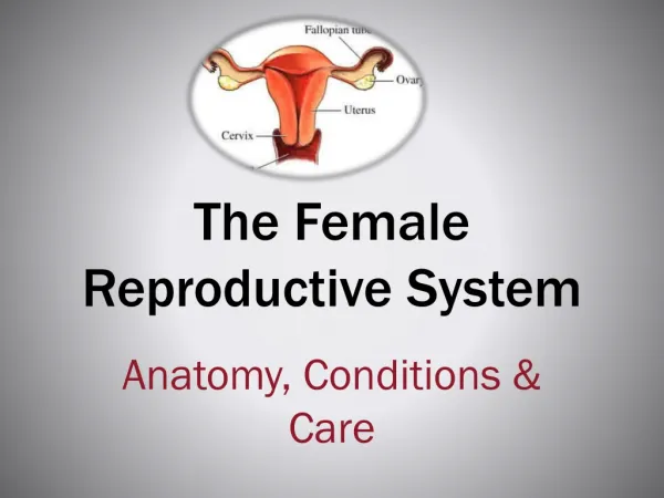
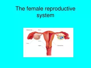
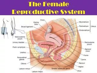
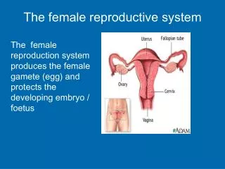
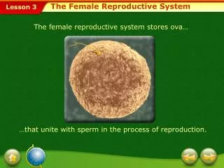
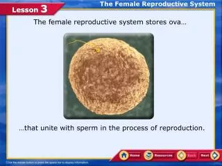
The reproduction system in females includes the ovaries and the uterus. Ovariesare the female gonads that produce the ova or eggs. Release of ova from ovaries is called ovulation
Uterusis the cavity in females, which is responsible for maintaining the fetus during its development and expelling it at the end of pregnancy. The uterus consists of two main layers. An outer muscle layer which is called myometrium, and an inner one, which is called endometrium andcontains numerous blood vessels and glands.
The uterus is connected to each ovary by the oviduct. Each oviduct picks up the ovum after ovulation serves as the site for fertilization of the ovum lets the ovum pass into the uterus
Fertilization (or conception) • The process in sexual reproduction that involves the union of sperm and ovum. To accomplish this, the female reproductive system accepts the sperm and transports it along with the released (from the ovaries) ovum to the oviduct for union. • The product of fertilization is known as an embryo.
Fertilization (or conception) Adhesion(Implantation) of the developing embryo to the wall of endometrium. This is the very early stage of pregnancy.
After two months the tissues have been differentiated and the developing living being is recognizable as human and is known as a fetus.
Fertilization, implantation and pregnancy do not take place without prior ovulation and appropriate preparation of the endometrium to accept the fertilized ovum Ovulation and appropriate preparation of the endometrium to accept the fertilized ovum take place during the menstrual cycle of the female.
Menstrual cycle • Menstruus means “monthly” • The menstrual cycle is a monthly repetitive series of changes a woman's body goes through to prepare for pregnancy, involving: • 1) release of the ovum from the ovary • 2) secretion of female sex hormones • 3) changes in the endometrium • Each cycle is characterized by changes that take place : • 1) in the ovaries and represent the ovarian cycle • 2) in the uterus and represent the uterine cycle. • If fertilization occurs, the cycles are interrupted and the female system adapts to nurture and protect the newly conceived human. • In the absence of sperm cells, fertilization does not occur and the cycle repeats. • The menstrual cycle begins after puberty and it is finally terminated by menopause.
Oogenesis (production of ova or eggs) before birth Mitotic divisions increase the number of the undifferentiated germ cells in the fetal ovaries, which are called oogonia Oogoniabegin the early steps of the first meiotic division but do not complete it. The produced cells, which are immature ova, are called primary oocytes. The primary oocytes remain in this state of meiotic arrest for years
Oogenesis (production of ova before birth) Each primary oocyte is surrounded by a single layer of cells, which are called granulosa cells. Together, an oocyte and the surrounding granulosa cells make up a primary follicle. Primary follicles (around2 million) serve as a reservoir from which all ova throughout the reproductive life of a female arise.
Oogenesis at puberty and after puberty • Of the original total pool of follicles at birth, about 300,000 remain at puberty, and only about 400 will mature and release ova, whereas the rest of them degenerate and die. • In each menstrual cycle only one primary developing oocyte reaches maturity and is ready for ovulation and fertilization. • By menopause, which occurs around the age of 50, the remaining few primary follicles either have already released their ova or degenerated. From this point on, the woman’s reproductive capacity ceases.
After puberty How does the primary oocyte develop to maturity in each menstrual cycle?
Just before ovulation • The primary oocyte which has been in meiotic arrest for years, completes its first meiotic division. • This division yields two daughter cells from a primary oocyte. • Only one of the daughter cells contains almost all the cytoplasm of the primary oocyte. This daughter cell is called secondary oocyte. • The secondary oocyte is destined to become the mature ovum. • The other daughter cell is called first polar body anddegenerates.
How does the primary oocyte develop to maturity in each menstrual cycle? • The final division in the secondary oocyte never completes, unless it is fertilized. • Sperm entry into the secondary oocyte is needed to trigger the second meiotic division. • The second meiotic division gives the second polar body, which degenerates and dies and the mature ovum, which contains almost all the cytoplasm of the secondary oocyte.
After puberty What happens to the follicle which contains the developing oocyte?
Ovarian cycle • The ovarian cycle is characterized by changes that take place in the ovaries and its follicles, including the divisions of oocytes. • The ovarian cycle consists of alternating follicular and luteal phases. • The luteal phase, is characterized by the presence of a yellow body, which is called corpus luteum • The follicular phase is characterized by the presence of developing follicles
Follicular phase Luteal phase The secretion of FSH, LH estrogen and progesterone differs between the follicular and luteal phases
Follicular phase • Only a few follicles (about 15-20 ) enter the follicular phase and they are called preantralfollicles. They came from primary follicles after proliferationofgranulosa cells and differentiation of the cells, which surround the granulosa cells, into theca cells. • The granulosa cells and the theca cells of the preantralfollicles are called follicular cells. • The oocyte in these follicles is the primary oocyte
The levels of FSH rise at the beginning of the follicular phase A few follicles enter the follicular phase
The preantral follicles increase in size and they are converted into antral or secondary follicles. The secondary follicles contain the antrum, which is a fluid-filled cavity in the middle of the granulosa cells. The antrum continues to expand as the secondary follicle grows
The preantral follicles increase in size and they are converted into antral or secondary follicles, under the influence of FSH.
Synthesis and secretion of estrogen by the follicles is stimulated by LH and FSH The secondary follicles are able to secrete estrogen, which, along with FSH and LH (secreted from the anterior pituitary) are required for further follicular development and antrum formation. As the follicle grows, estrogen is produced in increasing quantities.
Synthesis and secretion of estrogen by the follicles is stimulated by LH and FSH, which are secreted from the anterior pituitary
The secreted estrogen inhibitsthe secretion of FSH from the anterior pituitary by acting directly on the anterior pituitary and indirectly by inhibiting the secretion of GnRH from the hypothalamus, through a negative feedback loop. FSH secreted from the anterior pituitary is also inhibited by the action of inhibin, which is secreted by the follicular cells.
All the above result in a decrease of plasma FSH levels as the estrogen levels rise during the follicular phase.
The decrease in FSH secretion brings about degeneration (atresia) of all developing follicles except one (the most mature of the developing follicles), which is the dominant follicle and will grow to maturation.
The relatively low levels of estrogen also exert inhibitory effects on LH secretion (indirectly) by inhibiting the GnRH secretion from hypothalamus. The relatively low levels of estrogen during the follicular phase alone cannot completely suppress LH secretion. Complete suppression of LH secretion is achieved byprogesterone. Progesterone, like estrogen is a steroid hormone, which does not appear in the follicular phase.
The inability of the estrogen alone to completely inhibit LH secretion from anterior pituitary results in a slow increase of circulating LH levels. This increase of LH levels results in an increase of estrogen levels.
In this hormonal environment, the “dominant” follicle, grows more rapidly than the others and its antrum is largely expanded, and develops into the mature (preovulatory, tertiary, or Graafian)follicle.
The maturation takes place within about 14 days after the onset of follicular development. The oocyte in the follicle is still the primary oocyte. It is developed into the secondary oocyte just before ovulation.
The primary oocyte in the follicle is developed into the secondary oocyte just before ovulation, under the influence of high levels of LH that came from an abrupt, massive increase in LH secretion (LH surge). LH surge triggers ovulation
2 At this point and after the high levels of estrogen stimulate specifically the LH secretion from the anterior pituitary through a positive feedback loop. 1 The secretion of estrogen rises continuously under the influence mostly of LH, until it reaches a peak.
High levels of estrogen also increase FSH secretion, by increasing the secretion GnRH from the hypothalamus through a positive feedback loop • These levels are not as high as LH levels due to inhibition of FSH secretion by inhibin
The LH surge, whichlasts for about a day at midcycle triggers the production of local prostaglandins • Prostaglandins induce changes that rupture the follicle and promote ovulation.
The LH surge also causes the differentiation of the follicular cells into luteal cells and the formation of the corpus luteum. The formation of the corpus luteum automatically follows ovulation.
This is the starting point of the luteal phase of a menstrual cycle, which is characterized by the presence of the corpus luteum. The corpus luteum is yellow because it contains lipids rich in cholesterol.
The luteal cells secrete estrogen and progesterone. Progesterone is secreted at much larger quantities than estrogen
DIFFERENCE Progesterone dominates in the luteal phase Estrogen dominates in the follicular phase
As the luteal phase proceeds, the levels of estrogen and progesterone sharply decrease
As the luteal phase proceeds, the levels of estrogen and progesterone sharply decrease, because: • Progesterone (which dominates the luteal phase) acts on the hypothalamus and the anterior pituitary and inhibits the secretion of LH and FSH from the anterior pituitary • In this hormonal environment the corpus luteum degenerates and stops producing and secreting estrogen and progesterone.