Download
1 / 44
500 likes | 719 Views
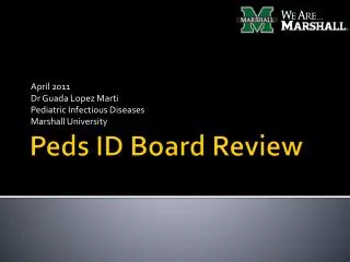
April 2011 Dr Guada Lopez Marti Pediatric Infectious Diseases Marshall University. Peds ID Board Review. Question 1.

E N D
April 2011 Dr Guada Lopez Marti Pediatric Infectious Diseases Marshall University Peds ID Board Review
Question 1 • A 5-year-old boy has been limping for 1 day and has a temperature to 39.0°C. Today he refuses to bear weight on his right leg. He is otherwise healthy, and his immunizations are up-to-date. He has not traveled outside of the US and has had no penetrating trauma, rash, or insect bites. On physical examination, the slightly ill child refuses to move his right leg. His thigh is tender and swollen proximal to his right knee. CRP= 12.5 mg/L (1.25 mg/dL) and the ESR =45 mm/hr. The next day, the blood culture shows no growth. The child remains febrile, with continued pain and erythema over the middle of the right thigh. The CRP is 1.4 mg/dLand the ESR is 85 mm/hr. • Of the following, the radiographic test that is MOST likely to establish the diagnosis and the need for surgical drainage in this child is: • A.computed tomography (CT) scan • B.magnetic resonance imaging (MRI) • C.plain radiograph of the lower extremities • D.radionuclide bone scan • E.tagged nuclear leukocyte (white blood cell) scan
Answer 1: B=MRI • Osteomyelitis is an infection of bone and marrow, most commonly a bacterial infection in immunocompetent children, whose pathogenesis usually is via hematogenous spread. Microorganisms also can be introduced into bone by direct inoculation (usually traumatic but also from surgery) and direct extension from a contiguous focus of infection. The incidence is highest in the first 2 decades of life, with approximately 25% of infections occurring in children younger than 2 years of age and 50% occurring in children younger than 5 years of age. • Radiographic studies, such as plain radiography, MRI, CT scan, bone scan, and leukocyte (tagged white cell) scan, frequently are helpful diagnostic tools in the evaluation of bone infection. However, MRI is the best imaging study for diagnosis of osteomyelitis and guiding management decisions • A conventional radiograph typically appears normal at the presentation of pain and fever because a reduction of greater than 50% in bone density must occur to detect a change. MRI provides specific anatomic information that is superior to CT scan or plain radiography . MRI can delineate subperiosteal or soft-tissue collections of pus.
Question 2 • You are asked to evaluate a 3-day-old infant who has developed thick drainage from both eyes. On physical examination, the infant has significant eyelid edema bilaterally and profuse purulent conjunctival drainage (Fig. 1). He was born at home and did not receive ocular prophylaxis at birth. A Gram stain of the conjunctival drainage reveals intracellular gram-negative diplococci (Fig. 2).
Figure 1: Bilateral edema and profuse purulent conjunctival drainage. Courtesy of Red Book Online.® 2009.
Figure 2: Gram stain of conjunctival drainage. Courtesy of Red Book Online.® 2009.
Question 2: cont • Of the following, the MOST appropriate antimicrobial therapy for this infant is: • A. ocular erythromycin ointment • B. oral erythromycin • C.parenteralampicillin and gentamicin • D.parenteralceftriaxone • E.parenteralceftriaxone and ocular erythromycin ointment
Answer 2: D parenteralceftriaxone • Ophthalmianeonatorum refers to conjunctivitis that occurs in the first 4 weeks after birth. It may be caused by chemical irritation; organisms transmitted from an infected maternal genital tract to the infant, such as Neisseriagonorrhoeae, Chlamydia trachomatis, and herpes simplex virus (HSV); or other bacterial organisms that may be transmitted prenatally or postnatally • Gonococcalophthalmia is the most serious cause because of its propensity to cause conjunctival scarring and permanent visual impairment. • Gonococcalophthalmia was common in the US prior to the widespread use of ocular prophylaxis and remains prevalent in developing countries, where ocular prophylaxis use is limited • Gonococcalophthalmia occurs most frequently 2 to 7 days after birth and is characterized by edema of the eyelid and a profuse purulent conjunctivalexudate, as described for the infant in the vignette
Management of gonococcalopthalmia • Neonates should be hospitalized and evaluated for disseminated infection. Initially, they should receive eye irrigation with saline solution hourly and then every 2 to 3 hours until the ocular discharge is eliminated, which may take several days, even with appropriate antimicrobial therapy. • Systemic antimicrobial therapy is required, and the treatment of choice is ceftriaxone, administered as a one-time intravenous or intramuscular dose. A longer course of therapy is recommended if disseminated infection is suspected or documented. Cefotaxime, rather than ceftriaxone, is recommended for infants who have hyperbilirubinemia. • Although ocular erythromycin therapy is effective in preventing gonococcal conjunctivitis, topical therapy is ineffective in the treatment of established infection and does not prevent disseminated disease. Oral erythromycin is ineffective in the treatment of gonococcal conjunctivitis. • Additional topical therapy is unnecessary and not recommended when systemic therapy is used.
Question 3 • You are evaluating a 2-year-old girl who has been in the US for 2 weeks after being adopted from a large, rural, crowded orphanage in Cambodia. She was abandoned at a bus station at about 1 month of age and has lived in the orphanage her entire life. No information is known about her biologic parents. • She received hepatitis B vaccine at 6 and 9 months. Hepatitis B serology performed at 9 months and 20 months of age showed hepatitis B surface antigen (HBsAg)–positive and hepatitis B surface antibody (HBsAb)–negative. • On physical examination, the small, thin toddler sits happily in her adoptive mother’s lap. Her height, weight, and head circumference are all far below the 5th percentile for age, with her height and weight at the 50th percentile for a 10-month-old child. She has a mildly distended abdomen without tenderness to palpation or hepatosplenomegaly
Question 3 cont-Her labs show: • Peripheral white blood cell count, 14x103/mcL (14x109/L) with 50% segmented neutrophils, 40% lymphocytes, and 10% monocytes • Hemoglobin, 9.5 g/dL (95 g/L) • Platelet count, 350x103/mcL (350x109/L) • Urinalysis, normal • Alanineaminotransferase, 135 units/L • Aspartateaminotransferase, 160 units/L • Albumin, 2.5 g/dL (25 g/L) • Alkaline phosphatase, 440 units/L • Hepatitis A immunoglobulin G (IgG), positive • Hepatitis C antibody, negative • HBsAg, positive • Antibody to HbsAg, negative • Hepatitis B virus early antigen, positive • IgM hepatitis B core antigen, negative
Question 3 cont • Of the following, the MOST likely diagnosis for this patient is: • A.acute hepatitis A infection • B.acute hepatitis B infection • C.chronic hepatitis B infection • D.no active hepatitis B infection • E.resolved hepatitis B infection
Answer 3: C chronic hepatitis B infection • The presence of: • Hepatitis B surface antigen (HBsAg) may indicate acute or chronic infection • Antibody to HBsAg (anti-HBs) identifies people who have resolved infection or who have been immunized • Hepatitis B e antigen (HBeAg) identifies infected people at increased risk for transmitting HBV • Antibody to hepatitis B core antigen (anti-HBc) identifies people who have acute, resolved, or chronic HBV infection • IgM antibody to HBcAg (IgM anti-HBc) identifies people who have acute or recent HBV infection
Question 4 • You are evaluating a 7-year-old girl whose mother is concerned about a foul-smelling discharge on the girl’s underwear that has been present for 3 days. The child also has complained about dysuria and urinary frequency. Findings on abdominal examination of the well-appearing prepubertal girl are unremarkable. Her genital examination reveals erythematous vulva and vagina with profuse yellow discharge. • Of the following, the pathogen that, if isolated, is MOST diagnostic of sexual abuse in this child is: • A.Gardnerellavaginalis • B.herpes simplex virus • C.Neisseriagonorrhoeae • D.Trichomonasvaginalis • E.Ureaplasmaurealyticum
Answer 4: C: N. gonorrhoeae • Infection with a sexually transmitted pathogen should be excluded in all children who present with clinical features suggestive of infection of the genital tract. The girl described in the vignette has symptoms and signs of acute vulvovaginitis. Although nonspecific infections due to overgrowth of bacterial flora is the most common cause of vulvovaginitis in this age group, evaluation should include diagnostic testing to exclude STDs. Isolating an infectious pathogen that is transmitted primarily through sexual contact has important social, medical, and legal implications. • The detection of certain sexually transmitted pathogens is diagnostic of sexual abuse in prepubertal children. These pathogens are N.gonorrhoeaeand C. trachomatisas well as T.pallidum, HIV, and hepatitis B and C viruses not acquired perinatally or via transfusion. • Some sexually transmitted pathogens, such as HSV and HPV also can be acquired through nonsexual contact. Genital HSV infection can occur through autoinoculation from oral lesions in children, and anogenital warts may result from perinatal transmission of HPV in children younger than 3 years of age. • However, sexual abuse should be suspected and investigated in all children who present with genital HSV or HPV infection.
Question 5 • A fellow in adolescent medicine contacts you to inquire about confirmation of the diagnosis of bacterial vaginosis (BV) with the DNA gene probe for detection of Gardnerellavaginalis. She is evaluating a 15-year-old sexually active girl in the adolescent clinic who presented with the complaint of 4 days of a milky, homogeneous vaginal discharge. The girl is otherwise well and has no fever or abdominal or pelvic pain. The fellow has prepared a wet mount stain from vaginal secretions
Question 5 • Of the following, the diagnosis of BV is confirmed BEST with: • A.bacterial culture • B.detection of G vaginalis by DNA probe • C.fruity odor detected on whiff test • D.Gram stain • E.vaginal pH greater than 4.5
Answer 5: E vaginal pH greater than 4.5 • BV, earlier designated nonspecific vaginosis, is the most common cause of vaginal discharge in females of childbearing age, accounting for 40% to 50% of cases. The absence of inflammation is the basis of the term "vaginosis" rather than "vaginitis." • BV likely is a synergistic infection caused by a complex alteration in the vaginal microbiologic flora with G vaginalis. The detection of G vaginalis alone by DNA probe, Gram stain, or culture is not diagnostic of BV because G vaginalis can be found in the anorectal flora of healthy adolescents of both sexes as well as that of asymptomatic, healthy infants and children. It is also part of the endogenous vaginal flora in women of reproductive age.
BV cont • In prepubertal children, the absence of lactobacilli is physiologic, thereby often making the diagnosis of BV difficult. A delicate balance of flora exists in the vagina, which can be disturbed easily by a number of factors that allow pathogens to develop more easily. • During childhood, infection typically is primarily in the vulva, with the vagina affected secondarily, a relation that generally is reversed during/after puberty. Prepubertal females are particularly susceptible to vulvovaginitis because of anatomic, physiologic, and hygienic considerations. The relative unprotected location of the vaginal introitus and its proximity to the anus, the lack of estrogen-induced mucosal cornification, and the neutral-to-alkaline pH of the vagina contribute to susceptibility
Question 6 • A 10-month-old infant develops a temperature to 40.0°C. After fever reduction, physical examination shows a nontoxic, well-appearing infant whose only finding of note is mild pharyngeal erythema. His immunizations are up to date. Laboratory studies reveal a white blood cell count of 17.0x10³/mcL (17.0x10⁹/L) with 65% neutrophils and normal urinalysis results. • Of the following, the MOST likely etiologic agent is • A.Adenovirus • B.cytomegalovirus • C.Haemophilusinfluenzae • D.Salmonella • E.Streptococcuspneumoniae
Answer 6: A.Adenovirus • Occult bacteremia is a syndrome in which a febrile patient who appears nontoxic and has no identified source of infection is found to have a positive blood culture. • This entity has been observed almost exclusively in children 3 though 36 months of age who have temperatures of 39.0°C or higher. Prior to the availability and routine administration of conjugate vaccines against Streptococcus pneumoniaeand Haemophilusinfluenzae type b in 2000 and 1990, respectively, as many as 7.7% of such children had bacteremia. • S pneumoniae and H influenzaetype b comprised approximately 65% and 17% of cases, respectively. Other reported causes of occult bacteremia included Salmonella, Neisseriameningiditis, and Staphylococcus aureus. Most highly febrile young children did not have bacteremia and were presumed to have a viral cause of the fever.
Answer 6 cont • With routine immunization of infants against H influenzaetype b and S pneumoniae, invasive H influenzaetype b infections have been all but eliminated in the United States, and the incidence of pneumococcal bacteremia has been reduced by 80% to 90%. • Occult bacteremia currently is a rare cause of fever in young children, and the infant described in the vignette is unlikely to have bacteremia, even with a total leukocyte count elevated to 17.0x10³/mcL (17.0x10⁹/L). In fact, it is not standard of care to obtain a blood culture in a young, well-appearing child, such as the infant described in the vignette. A blood culture demonstrating growth in such a child is more likely to be due to a contaminant than a pathogen
Question 7 • A 16-year-old boy presents to the emergency department with a 2-day history of dysuria and a urethral discharge. He denies testicular pain, arthritis, or conjunctivitis. Physical examination reveals a profuse purulent urethral discharge. Urinalysis shows the presence of leukocyte esterase and 25 white blood cells (WBCs)/high-power field (hpf). You obtain a Gram stain (Figure) of the urethral discharge.
Question 7 cont • Of the following, the MOST appropriate treatment for this adolescent is: • A.azithromycin • B.azithromycin and cefixime • C.ceftriaxone • D.ceftriaxone and metronidazole • E.ofloxacin
Answer 7: B azithromycin and cefixime • Clinical manifestations of urethritis may include dysuria, urinary frequency, urethral discharge, or urethral pruritus, although asymptomatic infections are common. It is important to document the presence of urethritis, as for the boy described in the vignette, which can be accomplished by the presence of any of the following: • A mucopurulent or purulent urethral discharge • The presence of leukocyte esterase in a male without urinary tract disease that could cause pyuria • At least 5 WBCs/hpf on a urethral smear Gram stain • A first voided urine with at least 10 WBCs/hpf • Infectious causes of urethritis in males frequently are categorized as those caused by Neisseriagonorrhoeae or those caused by nongonococcal agents (nongonoccalurethritis [NGU]). • Nongonoccalagents are the most common causes of urethritis. The most common cause of NGU is Chlamydia trachomatis, accounting for 30% to 50% of cases
cont • Because patients who have gonorrhea frequently are coinfected with chlamydia, all patients treated for gonococcal infection, including the boy in the vignette, also should be treated routinely with a regimen active against C trachomatis infection. • Appropriate therapy for gonorrhea is a single oral dose of cefixime or a single intramuscular injection of ceftriaxone. Nongonococcal causes of urethritis can be treated with a single oral dose of azithromycin or a 7-day course of doxycycline. • A single dose of azithromycin is preferred for NGU in adolescents because of adherence issues.
Question 8 • During a period of cefotaxime shortage, a pediatrician requests your advice on empiric antibiotic therapy for suspected sepsis in a 25-day-old infant who has morphologic features suggestive of possible DiGeorge syndrome. The baby had hypocalcemictetany in the immediate newborn period. On physical examination, the baby has minimal skin icterus, but no rash or petechiae. Findings on a complete blood count are within normal limits, and results of serum electrolytes assessment and a T-cell profile are pending. • Of the following, a CONTRAINDICATION to the use of ceftriaxone in this baby is the: • A. possible need to administer intravenous calcium • B. possible risk of seizures • C. presence of mild jaundice • D. risk of Candida infection • E. risk of ceftriaxone-induced hemolysis
Answer 8: A possible need to administer intravenous calcium • Cephalosporins are used widely in pediatrics, in part because adverse effects are infrequent, occurring at overall rate of about 1%. However, certain potential adverse reactions necessitate caution in using these antibiotics. • The infant described in the vignette has a history of tetany, and if hypocalcemia recurs, will require intravenous calcium solutions. One adverse effect associated with ceftriaxone use is a fatal reaction with ceftriaxone-calcium precipitates in lungs and kidneys among neonates who concomitantly receive intravenous calcium-containing products
cont • For this reason, ceftriaxone is contraindicated for infants younger than 28 days of age if they are receiving or are expected to receive calcium-containing products. • In addition, the high degree of protein-binding by ceftriaxone, which accounts for its long half-life, displaces bilirubin from albumin and, therefore, can increase the risk of hyperbilirubinemia and kernicterus in neonates. • The presence of jaundice, especially if physiologic, is only a relative contraindication, although it is another reason that ceftriaxone is not part of a standard empiric regimen for infants younger than 4 weeks of age. Ceftriaxone is not likely to increase the risk of seizures, Candida infection, or hemolysis in this baby.
Question 9 • A 15-day-old term male infant presents for evaluation of fever, fussiness, and decreased oral intake. He was born by precipitous normal spontaneous vaginal delivery to a 20-year-old woman who had no prenatal care. The infant was discharged from the hospital with his mother on postnatal day 3. • His mother has a history of using IV drugs during the pregnancy and reports being diagnosed with Chlamydia, gonorrhea, and syphilis in the past. • On physical examination, the fussy infant has a temperature of 38.9°C and a weight of 2.5 kg. His anterior fontanelle is full, he has nasal congestion with rhinorrhea, and scattered small and moist papules are evident on his lips and left buccal mucosa (Figure)
Question 9 cont… • His abdomen is distended, with a liver that is palpable 4 cm below the right costal margin and a spleen that is 3 cm below the left costal margin. There is a diffuse maculopapular, scaly, pink rash on his trunk, back, extremities, palms, and soles. Laboratory data show: • Peripheral white blood cell count of 4.0x10³/mcL (4.0x10⁹/L) • Hemoglobin of 10 g/dL (100 g/L) • Platelet count of 90x103/mcL (90x109/L) with 40% polymorphonuclear leukocytes, 55% lymphocytes, and 5% monocytes • Cerebrospinal fluid (CSF) parameters include: • Glucose of 55 mg/dL • Protein of 180 mg/dL • 2 red blood cells • 75 white blood cells
Question 9 cont… • Gram stain of the CSF shows no organisms. Blood, urine, and CSF specimens are sent for culture. Of the following, the test that is MOST likely to make the diagnosis in this patient is : • A. bacterial culture of body fluids • B. flow cytometry • C. polymerase chain reaction test of CSF • D. rapid plasma reagin test • E. viral culture of urine
Answer 9: D • Rapid plasma reagin test (RPR) • Case of congenital syphilis
Question 10 • A 4-year-old child presents with a 3-day history of fever, headache, and leg aches (myalgias) 2 weeks after a camping trip with her family in the southeastern US. On physical examination, you note a faint macular, blanching rash on her hands, feet, forearms, and trunk that is not raised is non-pruritic. The child is ill-appearing but nontoxic. Her neck is supple, findings on cardiovascular examination are normal, and she has a mildly diffusely tender abdomen. Both liver and spleen tips are palpable. A complete blood count shows a white blood cell count of 7.8x10³/mcL, with 15% bands, 80% neutrophils, and 5% lymphocytes; hemoglobin of 12 g/dL; and platelet count of 75x103/mcL. Of the following, the MOST appropriate next diagnostic test(s) is (are): • A. acute and convalescent serology • B. culture of blood or tissue biopsy • C. polymerase chain reaction testing • D. Proteus vulgarisOX-19 and OX-2 agglutination and the Weil-Felix serologic test • E. skin biopsy of the rash via direct immunofluorescence or immunoperoxidase staining
Answer 10: AAcute and convalescent serology • Diagnosis of RMSF
Question 11 • 16-year-old girl develops numbness, tingling, and pain in both feet that progresses proximally. Over the next several days, her lower extremities become weaker and she has difficulty walking. On physical examination, the afebrile adolescent has absent reflexes bilaterally in her upper and lower extremities and decreased muscle strength in her lower extremities. Examination of her CSFshows no white blood cells, protein of 63 mg/dL, and glucose of 60 mg/dL. She reports that she had a diarrheal illness 12 days before the onset of her symptoms. Of the following, the MOST likely pathogen associated with her diagnosis is: • A. Campylobacter jejuni • B. Escherichia coli type O157:H7 • C. Salmonella typhimurium • D. Shigellasonnei • E. Yersiniaenterocolitica
Answer 11: ACampylobacter jejuni • Guillain-Barre syndrome (GBS)after C. jejuniinfection • C jejuniinfection is the most commonly identified precipitant of GBS • The cardinal clinical features of GBS are progressive, fairly symmetric muscle weakness accompanied by absent or depressed deep tendon reflexes • Patients usually present a few days to a week after onset of symptoms. • The weakness can vary from mild difficulty with walking to nearly complete paralysis of all extremity, facial, respiratory, and bulbar muscles