Download
1 / 42
420 likes | 658 Views
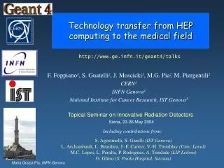
Technology transfer from HEP computing to the medical field. http://www.ge.infn.it/geant4/talks. F. Foppiano 3 , S. Guatelli 2 , J. Moscicki 1 , M.G. Pia 2 , M. Piergentili 2 CERN 1 INFN Genova 2 National Institute for Cancer Research, IST Genova 3.

E N D
Technology transfer from HEP computing to the medical field http://www.ge.infn.it/geant4/talks F. Foppiano3, S. Guatelli2, J. Moscicki1, M.G. Pia2,M. Piergentili2 CERN1 INFN Genova2 National Institute for Cancer Research, IST Genova3 Topical Seminar on Innovative Radiation Detectors Siena, 23-26 May 2004 Including contributions from: S. Agostinelli, S. Garelli (IST Genova) L. Archambault, L. Beaulieu, J.-F. Carrier, V.-H. Tremblay (Univ. Laval) M.C. Lopes, L. Peralta, P. Rodrigues, A. Trindade (LIP Lisbon) G. Ghiso (S. Paolo Hospital, Savona)
Technology transfer A real life case A dosimetric system for brachytherapy derived from HEP computing (but all the developments and applications presented in this talk are general) • Activity initiated at IST Genova, Natl. Inst. for Cancer Research (F. Foppiano et al.) • hosted at San Martino Hospital in Genova (the largest hospital in Europe) • Collaboration with San Paolo Hospital, Savona (G. Ghiso et al.) • a small hospital in a small town
precision • accurate model of the real configuration (from CT) • speed adequate for clinical use • user-friendly interface for hospital usage The goal of radiotherapy Delivering the required therapeutic dose to the tumor area with high precision, while preserving the surrounding healthy tissue Accurate dosimetry is at the basis of radiotherapy treatment planning Dosimetry system Calculate the dose released to the patient by the radiotherapy system
The reality Treatment planning is performed by means of commercial software The software calculates the dose distribution delivered to the patient in a given source configuration Open issues Cost Precision Commercial systems are based on approximated analytical methods, because of speed constraints Approximation in geometry modeling Approximation in material modeling Each treatment planning software is specific to one technique and one type of source Treatment planning software is expensive
Commercial factors • Commercial treatment planning systems are governed by commercial rules (as any other commercial product...) • i.e., they are produced and marketed by a company only if the investment for development is profitable Treatment planning systems for hadrontherapy are quite primitive not commercially convenient so far • No commercial treatment planning systems are available for non-conventional radiotherapy techniques • such as hadrontherapy • or for niche applications • such as superficial brachytherapy
Monte Carlo methods in radiotherapy Monte Carlo methods have been explored for years as a tool for precise dosimetry, in alternative to analytical methods de facto, Monte Carlo simulation is not used in clinical practice (only side studies) • The limiting factor is the speed • Other limitations: • reliable? • for “software specialists only”, not user-friendly for general practice • requires ad hoc modeling
Central-Axis depth dose Profile curves at 9.8 cm depth PLATO overestimates the dose at ~ 5% level Comparison with commercial treatment planning systems M. C. Lopes IPOFG-CROC Coimbra Oncological Regional Center L. Peralta, P. Rodrigues, A. Trindade LIP - Lisbon CT-simulation with a Rando phantom Experimental data with TLD LiF dosimeter CT images used to define the geometry: a thorax slice from a Rando anthropomorphic phantom
Bone Air A more complex set-up Beam plane Skull bone Tumor M. C. Lopes1, L. Peralta2, P. Rodrigues2, A. Trindade2 1 IPOFG-CROC Coimbra Oncological Regional Center - 2 LIP - Lisbon Head and neck with two opposed beams for a 5x5 and 10x10 field size An off-axis depth dose taken at one of the slices near theisocenter PLATO fails on the air cavities and bone structures andcannot predict accurately the dose to tissue that is surrounded byair Deviations are up to 25-30% In some tumours sites (ex: larynx T2/T3-stage) a 5% underdosage will decrease local tumour control probability from ~75% to ~50%
dosimetric system precise Develop a general purpose realistic geometry and material modeling with the capability of interface to CT images with a user-friendly interface low cost at adequate speed for clinical usage performing at
Requirements Calculation of3-D dose distributionin tissue Determination ofisodose curves Based on Monte Carlo methods Accurate description of physics interactions Experimental validation of physics involved Precision Accurate model of the real experimental set-up Realistic description of geometry and tissue Possibility to interface to CT images Simple user interface + Graphic visualisation Elaboration of dose distributions and isodoses Easy configuration for hospital usage Parallelisation Access to distributed computing resources Speed Transparent Open to extension and new functionality Publicly accessible Other requirements
Precision Based on Monte Carlo methods Accurate description of physicsinteractions Extension of electromagnetic interactions down to low energies (< 1 keV) Experimental validationof physics involved Microscopic validation of the physics models Comparison with experimental data specific to the brachytherapic practice
Code and documentation publicly distributed from web 1st production release: end 1998 2 new releases/year since then Developed and maintained by an international collaboration of physicists and computer scientists Run, Event and Track management PDG-compliant Particle management Geometry and Materials Tracking Detector response User Interface Visualisation Persistency Physics Processes
Atomic relaxation Fluorescence Auger effect Fe lines protons GaAs lines antiprotons shell effects e, down to 250 eV EGS4, ITS to 1 keV Geant3 to 10 keV Based on EPDL97, EEDL and EADL evaluated data libraries Based on Penelope analytical models Hadron and ionmodels based on Ziegler and ICRU data and parameterisations Barkas effect (charge dependence) models for negative hadrons ions Bragg peak
Validation Microscopic validation: verification of Geant4 physics Dosimetric validation: in the experimental context
Photon attenuation coefficient Stopping power 2N-L=13.1 – =20 - p=0.87 NIST Geant4-LowE Geant4-Standard 2N-S=23.2 – =15 - p=0.08 NIST Geant4-LowE Geant4-Standard Al Al proton straggling Microscopic validation many more validation results available! ions e-, Sandia database
Simulation Nucletron Data G. Ghiso, S. Guatelli S. Paolo Hospital Savona experimental mesurements F. Foppiano et al., IST Genova Distance along Z (mm) Dosimetric validation Comparison to manufacturer data, protocol data, original experimental data Ir-192 I-125
General purpose system For any brachytherapy technique Object Oriented technology Software system designed in terms of Abstract Interfaces For any source type Abstract Factory design pattern Source spectrum and geometry transparently interchangeable
Flexibility of modeling • Configuration of • any brachytherapy technique • any source type • through an Abstract Factory • to define geometry, primary spectrum Abstract Factory • CT DICOM interface • through Geant4 parameterised volumes • parameterisation function: material • Phantom • various materials • water, soft tissue, bone, muscle etc. General purpose software system for brachytherapy No commercial general software exists!
Realistic model of the experimental set-up Radioactive source Spectrum (192Ir, 125I) Geometry Patient Phantom with realistic material model Possibility to interface the system to CT images
Modeling the source geometry Precise geometry and material model of any type of source • Iodium core • Air • Titanium capsule tip • Titanium tube Iodium core I-125 source for interstitial brachytherapy Iodium core: Inner radius :0 Outer radius: 0.30mm Half length:1.75mm Titanium tube: Outer radius:0.40mm Half length:1.84mm Air: Outer radius:0.35mm half length:1.84mm Titanium capsule tip: Box Side :0.80mm Ir-192 source + applicator for superficial brachytherapy
Effects of source anisotropy Simulation Plato Simulation Plato Data Distance along X (mm) Distance along Z (mm) Effects of source anisotropy Plato-BPS treatment planning algorithm makes some crude approximation ( dependence, no radial dependence) Rely onsimulation for better accuracy than conventional treatment planning software Transverse axis of the source Comparison with experimental data Longitudinal axis of the source Difficult to make direct measurements
3-D view source Modeling the patient Modeling geometry and materials from CT data Modeling a phantom 3D patient anatomy Acquisition of CT image DICOM is the universal standard for sharing resources between heterogeneous and multi-vendor equipment file of any material (water, tissue, bone, muscle etc.) thanks to the flexibility of Geant4 materials package Geant4-DICOM interface developed by L. Archambault, L. Beaulieu, V.-H. Tremblay (Univ. Laval and l'Hôtel-Dieu, Québec)
User-friendly interface to facilitate the usage in hospitals Dosimetric analysis Graphic visualisation of dose distributions Elaboration of isodose curves Web interface Application configuration Job submission
Dose distribution Analysis of the energy deposit in the phantom resulting from the simulation Isodose curves Dosimetry Simulation of energy deposit through Geant4 Low Energy Electromagnetic package to obtain accurate dose distribution Production threshold: 100 mm 2-D histogram with energy deposit in the plane containing the source AIDA + Anaphe Python for analysis for interactivity Abstract Interfaces for Data Analysis + any AIDA-compliant analysis system
MicroSelectron-HDR source Leipzig applicator Dosimetry Endocavitary brachytherapy Dosimetry Interstitial brachytherapy Dosimetry Superficial brachytherapy Bebig Isoseed I-125 source
Application configuration Fully configurable from the web • Run modes: • demo • parallel on a cluster • (under test) • on the GRID • (under development) Type of source Phantom configuration # events
Speed adequate for clinic use Parallelisation Transparent configuration in sequential or parallel mode Access to distributed computing resources Transparent access to the GRID through an intermediate software layer
Performance Endocavitary brachytherapy 1M events 61 minutes Superficial brachytherapy 1M events 65 minutes Interstitial brachytherapy 1M events 67 minutes on an “average” PIII machine, as an “average” hospital may own Monte Carlo simulation is not practically conceivable for clinical application, even if more precise
DIANE prototype for an intermediate layer between applications and the GRID Transparentaccess to a distributed computing environment Parallelisation Access to the GRID DIANE DIstributed ANalysis Environment Hide complex details of underlying technology R&D in progress for Large Scale Master-Worker Computing http://cern.ch/DIANE Developed by J. Moscicki, CERN
preliminary: further optimisation in progress Performance: parallel mode on a local cluster Endocavitary brachytherapy 1M events 4 minutes 34’’ Superficial brachytherapy 1M events 4 minutes 25’’ 5M events 4 minutes 36’’ Interstitial brachytherapy on up to 50 workers, LSF at CERN, PIII machine, 500-1000 MHz Performance adequate for clinical application, but… it is not realistic to expect any hospital to own and maintain a PC farm
Running on the GRID • Via DIANE • Same application code as running on a sequential machine or on a dedicated cluster • completely transparent to the user A hospital is not required to own and maintain extensive computing resources to exploit the scientific advantages of Monte Carlo simulation for radiotherapy Any hospital – even small ones, or in less wealthy countries, that cannot afford expensive commercial software systems – may have access to advanced software technologies and tools for radiotherapy
Current #Grid setup (computing elements): 5000 events, 2 workers, 10 tasks (500 events each) - aocegrid.uab.es:2119/jobmanager-pbs-workq - bee001.ific.uv.es:2119/jobmanager-pbs-qgrid - cgnode00.di.uoa.gr:2119/jobmanager-pbs-workq - cms.fuw.edu.pl:2119/jobmanager-pbs-workq - grid01.physics.auth.gr:2119/jobmanager-pbs-workq - xg001.inp.demokritos.gr:2119/jobmanager-pbs-workq - xgrid.icm.edu.pl:2119/jobmanager-pbs-workq - zeus24.cyf-kr.edu.pl:2119/jobmanager-pbs-infinite - zeus24.cyf-kr.edu.pl:2119/jobmanager-pbs-long - zeus24.cyf-kr.edu.pl:2119/jobmanager-pbs-medium - zeus24.cyf-kr.edu.pl:2119/jobmanager-pbs-short - ce01.lip.pt:2119/jobmanager-pbs-qgrid Spain Greece Poland Portugal Traceback from a run on CrossGrid testbed Resource broker running in Portugal matchmaking CrossGrid computing elements
Extension and evolution • Configuration of • any brachytherapy technique • any source type System extensible to any source configuration without changing the existing code • General dosimetry system for radiotherapy • extensible to other techniques • plug-ins for external beams • (factories for beam, geometry, physics...) • treatment head • hadrontherapy • ... Plug-ins in progress
A medical accelerator for IMRT Build a simulation tool which determines the dose distributions given in a phantom by the head of a linear accelerator used for IMRT. Many algorithms were developed to estimate dose distributions, but even the most sophisticated ones resort to some approximations. These approximations might affect the outcome of dose calculation, especially in a complex treatment planning as IMRT. step and shoot IMRT generates tightly conforming dose distributions. This microscopic control allows IMRT to produce dose distribution patterns that are much closer to the desired patterns than possible previously
Work in progress... • The user can choose the energy and standard deviation of the primary particles energy distribution (Gaussian) • The primary particles (e-) leave from a point source with random direction (0˚< θ < 0.3˚) and a gaussian distribution • The head components modeled include: target, primary and secondary collimators, vacuum window, flattening filter, ion chamber, mirror, vacuum and air • Each pair of jaws can be rotated through an axis that is perpendicular to the beam axis • The actual analysis produces some histograms from which the user can calculate the Percent Depth Dose (PDD) and the flatness at the following depths in the phantom: 15 mm, 50 mm, 100 mm and 200 mm.
(very) Preliminary results Percent Depth Dose Flatness
GEANT4 simulation CATANA hadrontherapy talk by P. Cirrone on Monday Real hadron-therapy beam line
Dosimetry in interplanetary missions Aurora Programme vehicle concept Dose in astronaut resulting from Galactic Cosmic Rays
Conclusions precise, versatile, fast, user-friendly, low-cost dosimetry • Physics & software technology from HEP have a potential to address key issues in medical physics • The social impact of technology transfer from HEP computing may be significant • What is the support of HEP to technology transfer? Geant4 + AIDA/Anaphe/PI + WWW + DIANE + GRID =
Thanks! This project has fostered a collaborative aggregation of contributions from many groups all over the world • G. Cosmo (CERN, Geant4) • L. Moneta, A. Pfeiffer(Anaphe/PI, CERN) • J. Knobloch (CERN/IT) • S. Agostinelli, S. Garelli (IST Genova) • G. Ghiso, R. Martinelli (S. Paolo Hospital, Savona) • G.A.P. Cirrone, G. Cuttone (INFN LNS, CATANA project) • M.C. Lopes, L. Peralta, P. Rodrigues, A. Trindade (LIP Lisbon) • L. Archambault, J.F. Carrier, L. Beaulieu, V.H. Tremblay (Univ. Laval) the authors F. Foppiano (IST) – medical physicist S. Guatelli, M. Piergentili (Univ. and INFN Genova) – students J. Moscicki (CERN) – computer scientist M.G. Pia (INFN Genova) – particle physicist