Download
1 / 48
520 likes | 1.47k Views
Complex Regional Pain Syndrome. Case. 53 yo male w/ complaints of severe LLE pain Pain has been present for “a few years”, but the severity has increased significantly over the previous 8 months Described as sharp and burning, with areas of numbness and tingling

E N D
Case • 53 yo male w/ complaints of severe LLE pain • Pain has been present for “a few years”, but the severity has increased significantly over the previous 8 months • Described as sharp and burning, with areas of numbness and tingling • His foot is generally ‘dark red’ and often swollen • He is unable to wear socks because his pain is exacerbated by clothing touching his skin • He describes weakness in the extremity to the point that he occasionally falls to the ground • He reports a history of frequent stress fractures and sprains during his days in the marines. He underwent a left anke tri-fusion 3 years ago
Historical Perspective • During the Civil War, Silas Weir Mitchell observed a chronic pain syndrome in soldiers who suffered traumatic nerve injuries • Their symptoms included constant burning pain and significant trophic changes
He described this syndrome using the term causalgia (from the Greek kausis – “burning” and algos – “pain”)
Half a century later, a French surgeon named Rene Leriche implicated the sympathetic nervous system in causalgic pain. He treated these patients with surgical sympathectomy
In the 1950’s, John Bonica (founder of the IASP) introduced the phrase reflex sympathetic dystrophy after noticing the efficacy of temporary blockade of the sympathetic nervous system in these patients
There have since been many confusing terms used to describe the condition: • Acute atrophy of the bone • Algodystrophy • Algoneurodystrophy • Chronic traumatic edema • Postinfarctional sclerodactyly • Post-traumatic algodystrophy • Post-traumatic dystrophy • Post-traumatic osteoporosis • Post-traumatic spreading neuralgia • Post-traumatic sympathetic dystrophy • Pseudodystrophy • Reflex neurovascular dystrophy • Shoulder hand syndrome • Sudeck’s dystrophy • Sympathalgia • Traumatic angiospasm • Traumatic vasospasm
In 1993, the IASP introduced the term Complex regional pain syndrome to describe all pain states that previously would have been diagnosed as RSD or causalgia-like syndromes
CRPS • Complex: Varied and dynamic clinical presentation • Regional: Non-dermatomal distribution of symptoms • Pain: Out of proportion to the inciting events • Syndrome: Constellation of symptoms and signs
The term “sympathetic” was avoided in the revised definition because its contribution is not constant across patients • CRPS pain may be “sympathetically maintained pain” (SMP) or “sympathetically independent pain” (SIP)
CRPS can be separated into two types based on the presence or absence of a nerve injury • CRPS type I: A syndrome that develops after an initiating noxious event that may or may not be associated with a period of immobilization • CRPS type II: Differs from CRPS type I by the presence of a known injury to a nerve or nerves
Epidemiology • Incidence: 5.46/100,000/year • Prevalance: 20.57/100,000 • Female:Male ratio: 3-4:1 • 80-85% have experienced preceeding trauma (fractures, surgery) • 10% have experienced minor trauma • 5-10% occur spontaneously
There is no correlation between the severity of trauma and the degree of CRPS symptoms. • No psychological factor or personality structure predisposing for CRPS has been identified, however, studies have demonstrated that up to 80% of CRPS patients had experienced ‘stressful life events’ close to the time of diagnosis.
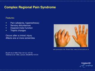
Clinical presentation • Characteristic triad of symptoms comprising autonomic, sensory, and motor disturbances • Triad can differ amongst individuals • Symptoms will generally change over time in a given individual
Distal edema – 80% • Skin temperature changes – 80% • The affected area is initially warm, but over the course of the disease the skin temp decreases • Skin color changes • Initially red, becomes pale in chronic disease • Altered sweating • Increased sweating more common • Nail and hair changes • Increased growth in early disease
Spontaneous pain: • Often described as burning, aching, throbbing, shooting, or deep pressure pain • Hyperpathia • Hyperalgesia • Allodynia • Motor changes: • Weakness, distal tremors, dystonia, myoclonus • Not clear whether these are part of the clinical presentation of the disease or a result of protection/disuse of the painful limb
Bony changes: • Osteoporosis – periarticular distribution • Joint stiffness
Patients often have associated psychological and psychiatric disturbances • These are generally consequences of the disorder rather than causes thereof
Pathophysiology • Three main hypotheses • Facilitated neurogenic inflammation • Autonomic dysfunction • Neuroplastic changes within the CNS
Neurogenic inflammation • Classic inflammatory signs are present in CRPS: pain, swelling, erythema, hyperthermia and impaired function • However, when clinical chemistry parameters for inflammation are evaluated, there are no differences between CRPS patients and controls • With neurogenic inflammation, distinct classes of C-fibers called mechano-heat-insensitive C-fibers have both an afferent function in the mediation of pain and itch as well as an efferent neurosecretory function, releasing neuropeptides via ‘axon reflex’ • Action potentials in these fibers can be conducted retrogradely to terminal branches via axon collaterals where neuropeptides such as substance P and calcitonin-gene-related peptide (CGRP) are released • Substance P provokes plasma protein extravasation (edema) and appears to have a role in osteoclastic activity • CGRP induces vasodilation (hyperthermia and erythema), increases sweating, and appears to be involved in hair growth
Autonomic dysfunction • Pathological sympatho-afferent coupling: Peripheral nociceptors develop adrenergic sensitivity (mainly alpha-2 receptors) such that tonic sympathetic efferent activity leads to their activation • Painful impulses via these nociceptors maintain the central nervous system in a sensitized state • Painful and non-painful stimuli to the affected limb result in hyperalgesia and allodynia, respectively • Catecholamine levels, however, are actually lower in the affected extremity, thus, it is not a problem of excessive sympathetic nerve output
Neuroplastic changes within the CNS • Studies using functional brain imaging in patients with CRPS have found a significant degree of cortical reorganization in the central sensory and motor cortices • The amount of reorganization positively correlates with the extent of pain intensity • The areas of reorganization were found to be reversible in adequately treated patients
Diagnosis • Based exclusively on the characteristic clinical features of the condition • IASP diagnostic criteria for Complex Regional Pain Syndrome: • Presence of an initiating noxious event or cause of immobilization • Continuing pain, allodynia, or hyperalgesia, with pain disproportionate to any inciting event • Evidence at some time of edema, changes in skin blood flow, or abnormal sudomotor activity in the region of pain • Diagnosis is excluded by the existence of conditions that would otherwise account for the degree of pain and dysfunction
More on the IASP diagnostic criteria: • Introduced in 1994 • High sensitivity, low specificity • Not empirically validated prior to introduction • Studies have demonstrated that only a minority of physicians strictly follow these recommendations
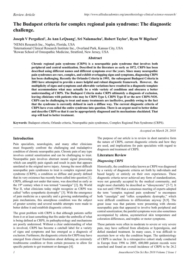
A modified diagnostic criteria was proposed by the IASP in 2007 to increase specificity (the ‘Budapest criteria’): • Continuing pain that is disproportionate to any inciting event • Must report at least one symptom in three of the four following categories: • Sensory: hyperalgesia, allodynia • Vasomotor: temp. asymmetry, skin color changes or asymmetry • Sudomotor/Edema: edema, sweating changes or asymmetry • Motor/Trophic: decreased range of motion, weakness, tremor, dystonia, trophic changes • Must display at least one sign at the time of evaluation in two or more of the following categories: • Sensory: hyperalgesia, allodynia • Vasomotor: temp. asymmetry, skin color changes or asymmetry • Sudomotor/Edema: edema, sweating changes or asymmetry • Motor/Trophic: decreased range of motion, weakness, tremor, dystonia, trophic changes • No other diagnosis better explains the signs and symptoms
Differential diagnosis: • Unrecognized local pathology (fracture, sprain) • Traumatic vasospasm • Cellulitis • Lymphedema • Raynaud’s disease • Thromboangiitis obliterans • Erythromelalgia • DVT • Also, nerve entrapment syndromes, occupational overuse syndromes, and diabetic neuropathy
Diagnostic tests • Three-phase bone scintigraphy: • Significant uptake in the metacarpophalangeal or metacarpal bones appears to have high sensitivity and specificity for CRPS • The best timing for this study is in the subacute (up to 1 year) phase of the condition
Plain radiographs, x-ray bone densitometry, and magnetic resonance imaging have not been shown to be sensitive or specific for CRPS
Tests used more in the research setting: • Quantitative sensory testing • May reveal impairment of warm and cold sensation and heat pain in patients with CRPS • Autonomic function testing • Infrared thermometry • Infrared thermography • Quantitative sudomotor axon reflex test • Thermoregulatory sweat test • Laser Doppler flowmetry
Diagnostic tests used to assess for a sympathetically maintained component: • Sympathetic ganglia blockade • Regional intravenous blockade with Guanethidine • Phentolamine infusion test
Sympathetic ganglia blockade • Stellate ganglion: upper extremities • Lumbar paravertebral ganglia: lower extremities **The results of these blocks need to be interpreted carefully** • Evaluation of the effects of the block on sudomotor and vasoconstrictor function by assessing skin blood flow, temperature and resistance is vitally important to know whether the sympathetic block is complete, especially in patients who do not experience significant pain relief. • Local anesthetic can spread to nearby nerve roots, resulting in somatic nerve block that may significantly affect the patient’s pain. Therefore, it is important to do a careful sensory examination of the affected area. • If a large dose of LA is used, systemic uptake may provide some pain relief. • The invasive procedure itself may have a significant placebo effect. • Visceral sensory afferent fibers traveling with the sympathetic chain may be blocked and result in pain relief.
Regional intravenous block with Guanethidine • Drug taken up by postganglionic sympathetic nerves where it depletes norepinephrine stores and prevents further release of norepinephrine for 1-2 days • The initial depletion of norepinephrine stores can cause short-term excitation of nociceptors due to the increased norepinephrine release leading to increased pain during the procedure
Phentolamine infusion test protocol: • Patient preparation • Informed written consent is obtained • A standardized set of directions is read to the patient; the patient is told that pain may increase, decrease, or stay the same, and that the results will help guide future treatments • The patient is placed in supine position • ECG, BP, HR, and skin temp are monitored • An IV is established • A baseline pain level is established (must be >4/10) • Saline pretreatment • Lactated Ringer’s solution is administered at 600 mL/hr throughout the test • Sensory testing is done every 5 min for at least 30 min or until a stable pain rating is achieved. If the pain level is not stable, the test is deferred
Phentolamine infusion test protocol (cont.) • Phentolamine infusion • Propranolol 1-2 mg is administered intravenously • An infusion of phentolamine (1 mg/kg) is given over a 10-min period in single-blinded fashion (no clues provided to the patient on time of initiation of drug infusion) • Sensory testing is continued every 5 min during phentolamine infusion • Post-phentolamine testing • Sensory testing is continued for 15-30 min • ECG, BP, HR, and skin temp monitoring are continued for 30 min or longer, depending on the stability of vitals and presence of orthostatic hypotension
Management of CRPS • No scientifically validated cure exists • Therapy is directed at managing the signs and symptoms of the disease • A multidisciplinary approach utilizing pharmacotherapy, physical therapy and psychological therapy is most appropriate
Pharmacologic therapy • Drugs demonstrated to be effective for CRPS based on randomized controlled trials, and their proposed mechanism of action: • Prednisone (oral): anti-inflammatory, neuronal membrane stabilizer • Vitamin C (oral): antioxidant • Alendronate (IV): osteoclast inhibitor • Bretylium (IV): Autonomic ganglia blocker • Ketansarin (IV): serotonin and alpha receptor antagonist • Phentolamine (IV): alpha-1 receptor antagonist • Lidocaine (IV): sodium channel blocker • DMSO (topical): free radical scavenger • Calcitonin (intranasal): osteoclast inhibitor • Clonidine (epidural): alpha-2 receptor agonist • Baclofen (intrathecal): GABA-B receptor agonist
Other medications with reports of variable efficacy include: • AED’s • TCA’s • SNRI’s • Ketamine • NSAID’s • Opioids
Physical/Occupational therapy • Early physical therapy is essential to avoid atrophy and contractures of the affected limb • PT/OT have been shown to reduce pain and motor impairment, and improve function and coordination ability of the limb • Requires that the patient take an active role in their care
Psychological therapy • An integral part of the multidisciplinary treatment approach. Many patients with CRPS have a significant amount of psychological dysfunction, which is a reflection of the disease process itself as opposed to a cause thereof. • Pain coping skills • Biofeedback • Relaxation training • Cognitive behavioral therapy • Mirror therapy
Invasive/Interventional therapy • Sympathetic nerve blocks • Both diagnostic and therapeutic for SMP • Useful for helping patients tolerate physical and occupational therapy • Effectiveness of repeat blocks may be unpredictable • If a plateau of responsiveness to these blocks is reached, more advanced interventional therapies may need to be considered
Spinal cord stimulation • Randomized controlled trials have demonstrated a significant reduction in level of pain and improvement in functional status and quality of life in patients with SCS plus physical therapy compared to those undergoing physical therapy alone. • A case series found significantly reduced pain intensity in patients with SCS at 6, 12, and 24 months after implantation • A follow up study reported a constant pain reduction and health-related quality of life improvement in these patients 2 years after implantation
Surgical/chemical sympathectomy • Surgical sympathectomy appears significantly more effective than chemical sympathectomy • One study of 73 patients that had undergone surgical sympathectomy reported a patient satisfaction rate of 77% • There is, however, considerable risk of developing a post-sympathectomy pain syndrome that may be the result of a denervation supersensitivity of alpha receptors • Peripheral nerve stimulator • Limited literature, but there are papers that report positive results in patients with CRPS • Intrathecal baclofen pump
Conclusion • CRPS is a complicated chronic pain syndrome with variable clinical presentation and complicated diagnostic criteria. Diagnosis is made on a clinical basis and treatment is best managed with a multidisciplinary approach including medications, interventional procedures, physical therapy and psychological therapy
References: • C Maihofner, F Seifert and K Markovic. Complex regional pain syndromes: new pathophysiological concepts and therapies. Eur J Neuro 2010, 17: 649-660 • Hsu. Practical management of complex regional pain syndrome. Am J Therap 2009, 16: 147-154 • QH Tran, S Duong, P Gertini, RJ Finlayson. Treatment of complex regional pain syndrome: a review of the evidence. Can J Anaesth. 2010, 57(2): 149-166 • M de Mos, MC Sturkenboom, FJ Huygen. Current understandings on complex regional pain syndrome. Pain Pract. 2009, 9(2): 86-89 • Benzon. Essentials of Pain Medicine and Regional Anesthesia. Chapters 46-47, 376-385 • Longnecker. Anesthesiology. Chapter 91, 2020-2041