Other Methods
Other Methods. Northern blotting – analyzes mRNA expression in tissue In situ hybridization – Visualizing gene activity (mRNA) directly in fixed cells or tissues Western blotting – analyzes proteins expressed in tissue

Other Methods
E N D
Presentation Transcript
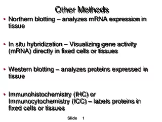
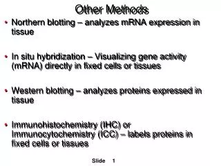
Other Methods • Northern blotting – analyzes mRNA expression in tissue • In situ hybridization – Visualizing gene activity (mRNA) directly in fixed cells or tissues • Western blotting – analyzes proteins expressed in tissue • Immunohistochemistry (IHC) or Immunocytochemistry (ICC) – labels proteins in fixed cells or tissues
Northern Blotting Purpose: Can tell us if a particular gene is being expressed in a specific cell or tissue type or if there are changes in levels of expression ----------------------------------------------------------------------------------------------------------------------------------- General Procedure: • Isolate mRNA from cells • Separate mRNA with gel electrophoresis • Blot (transfer) the mRNA to a membrane • Label mRNA you are interested in (probe with a complementary sequence that binds your mRNA of interest)
In situ Hybridization Purpose: Allows us to visualize gene activity directly in tissues or cells ----------------------------------------------------------------------------------------------------------------------------------- General Procedure: • Tissues may be cut into thin sections or entire embryo may be adhered to a microscope slide • mRNA probes are labeled with a dye or enzyme that makes a colored precipitate • Tissues are imaged with a microscope
Western Blotting Purpose: Detects levels of proteins being produced in a particular cell or tissue ----------------------------------------------------------------------------------------------------------------------------------- General Procedure: • Isolate proteins • Separate proteins with gel electrophoresis • Blot (transfer) the proteins to a membrane • Label protein you are interested in (antibody that binds specifically to your protein) • Bands can tell if protein is present and if there are different molecular forms of it
NE EX NE EX NE EX NE EX Not Breeding Not Breeding Turkey NB Turkey BR Breeding Breeding Female Male Zebra finch anterior pituitaries Example of Western Blotting Prolactin Isoforms in the Anterior Pituitary of Zebra Finches The lane on the extreme left is the ladder (mol wt marker). Tissue extracts in the remaining lanes are labeled with a prolactin antibody. Some birds demonstrate a second, slightly heavier band of prolactin. Because it is heavier, it migrates more slowly, and shows prolactin exists in two forms in these birds; the heavier prolactin has been modified by adding a sugar group.
Microscopy The idea behind any type of microscopy is to see inside the tissue and/or cell. What we see and how well we see it, depend upon: 1. Type of microscope 2. Tissue preparation 3. Tissue staining
Types of Microscopes Light Microscope: Resolution of about 0.2mm Good for routine staining Fluorescent microscope: Fluorescent dyes are excited by specific wavelength of light and re-emit it at higher wavelengths (lower energy). (ex: FITC – a very common fluorescent dye – is excited at 488, emits at 525nm - looks green) Filters eliminate stray light, so the image is sharper
Confocal Microscope Uses a laser to excite fluorescent dye in tissue or cell one spot at a time Precisely positioned pinholes allow only in-focus light to pass through Images in XYZ planes Computer takes all images and creates a 3D reconstruction
Confocal Microscope Confocal microscope: 0.2-0.5mm resolution 3D images can be obtained http://www.jneurosci.org/content/vol26/issue46/images/data/11870/DC1/Fig._12C-PBC-3D_animation-Ruangkittisakul_et_al.mov Ion movements can be recorded ]http://www.jneurosci.org/content/vol26/issue46/images/data/11870/DC1/Fig._12A-PBC_calcium_oscillations-Ruangkittisakul_et_al.mov Anterior pituitary of a zebra finch. Cells secreting prolactin appear red, while those secreting growth hormone appear green. An individual cell in the lower right corner has both hormones and appears yellow. The dark “hole” in the middle of the cell is its nucleus. From: Christensen, 2007
Tissue Preparation Fresh tissue is thick, mushy, and opaque. Also has active enzymes and bacteria that promote degradation. 3 basic steps: 1. Fixation – stabilizes structure 2. Embedding & sectioning – makes it hard so we can cut thin slices 3. Staining – provides contrast so we can see it easier
Histological Techniques Fixation: stabilizes structure stops enzymes prevents bacterial degradation (putrefaction) The most common fixatives cross-link tissue proteins. An example is formaldehyde.
Embedding & Sectioning Embedding: Infiltrate tissue with harder substance that is more easily cut into thin sections 1. Resin – plastic, used for very thin sections, 0.5-1µm 2. Paraffin – used for sections >5µm 3. Freezing – used for sections 20-100µm
Sectioning Paraffin (frozen similar) 1. Sectioned with arotary microtome 2. Metal or glass knife or razor blade 3. Sectionsto1-2 µm thick
Staining Staining - introduces contrast to tissue which is otherwise essentially transparent Common stains: Hematoxylin (looks blue-purple) & eosin (looks pink-red) Usually hematoxylin and eosin are used together (H&E)
Histochemistry Can stain DNA & visualize replicating cells
DNA BRd-U - bromodeoxyuridine - analog of thymidine, incorporated when DNA replicates a. Antibody stains labeled cells b. Can follow time sequence of cell division
Advantages 1. High specificity for molecular species 2. Can be used for light, confocal, or EM Disadvantages Time consuming & expensive 2. Fixation can interfere with Ab binding 3. Reproducibility - false positives - cross reactivity 4. Difficult to get Abs to small molecules Immunohistochemistry (IHC) Uses antibodies to detect specific molecules in tissue
Immuno methods 1. Direct method - label (dye) is on the antibody
Immuno methods 2. Indirect method - label is on a secondary antibody, amplifies label. Primary antibody binds to molecule of interest Second antibody (labeled with dye) against species in which primary was raised - eg, goat anti-rabbit antibody
Labels Fluorescent dyes - FITC, rhodamine, Cy3, Cy5 - different excitation and emission spectra allows double labeling Enzymes (horse-radish peroxidase (HRP) or alkaline phosphatase) give colored precipitate