Download
1 / 17
170 likes | 341 Views
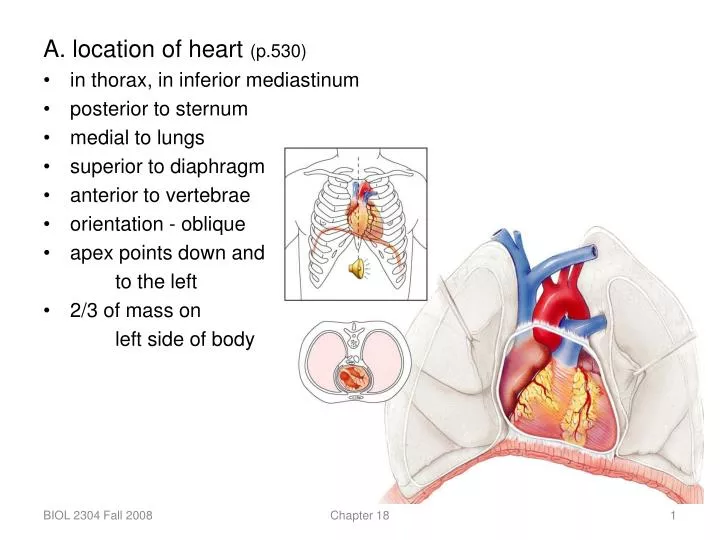
A. location of heart (p.530) in thorax, in inferior mediastinum posterior to sternum medial to lungs superior to diaphragm anterior to vertebrae orientation - oblique apex points down and to the left 2/3 of mass on left side of body.

E N D
A. location of heart (p.530) • in thorax, in inferior mediastinum • posterior to sternum • medial to lungs • superior to diaphragm • anterior to vertebrae • orientation - oblique • apex points down and to the left • 2/3 of mass on left side of body Chapter 18
B. overall function - creates a pressure gradient that moves blood through the vascular system (p.529) • perfusion = blood flow through tissues and organs • 1. left heart = systemic pump • supplies body tissues and returns • blood to heart • 2. right heart = pulmonary pump • takes blood to lungs to pick up oxygen • and get rid of carbon dioxide Chapter 18
C. pericardium – serous membranes of heart (p. 531) 1. parietal pericardium - double membrane surrounding heart, attached only at base of vessels on superior margin of heart, also called the pericardial sac a. outer layer (fibrous pericardium) = fibrous c.t. b. inner layer (parietal layer of serous pericardium) = serous membrane (simple squamous e. + areolar c.t.) 2. visceral [layer of serous] pericardium - attached to surface of heart = serous membrane (simple squamous e. + areolar c.t.) Chapter 18
D. heart wall (p. 531, 532) 1. epicardium = visceral pericardium 2. myocardium = cardiac m. ; bundles of cells arranged in spiral pattern 3. endocardium = simple squamous e. + areolar c.t. Chapter 18
E. chambers and vessels (p. 535) 1. atria (right and left) superior separated from each other by interatrial septum atrioventricular groove / coronary sulcus separates atria from ventricles Chapter 18
a. right atrium forms right border of heart • auricle = outer wall of atrium • fossa ovalis in interatrial septum • superior and inferior vena cavae return blood from body to heart • coronary sinus returns blood from coronary circulation to heart b. left atrium forms most of posterior surface • auricle • pulmonary veins return blood from lungs to heart Chapter 18
2. ventricles (right and left) inferior interventricular septum separates ventricles from each other interventricular sulci = groves on outside of heart over interventricular septum; anterior and posterior trabeculae carnae = ridges of muscle tissue seen on internal surface of ventricles Chapter 18
a. right ventricle (thinner wall) • right border of heart • pulmonary trunk carries blood from heart to lungs b. left ventricle (thicker wall) • apex and inferior surface of heart • aorta carries blood from heart to body Chapter 18
F. valves - control direction of blood flow; prevent backflow valves are opened and closed by pressure gradients (p. 539) Chapter 18
1. atrioventricular (AV) valves have cusps attached to ventricular wall by chordae tendineae at papillary muscles a. right = tricuspid b. left = bicuspid or mitral Chapter 18
2. semilunar (SL) valves have 3 cusps attached to wall at junction of ventricle and artery a. right = pulmonary b. left = aortic Chapter 18
G. fibrous skeleton (p. 538) • made of dense fibrous c.t. • separates atria and ventricles • surrounds and anchors valves; prevents overdilation • serves as insertion for cardiac muscle bundles • blocks electrical impulses Chapter 18
H. conducting system (p. 543) 1. autorhythmic cells vs contractile cells a. contractile cells (99%) are specialized for contraction b. autorhythmic cells (1%) are specialized for generating and conducting electrical signals smaller diameter, less actin and myosin than contractile cells automatically generate action potentials and conduct cardiac impulse through heart Chapter 18
2. conducting system (components red, conduction pathway blue, effect on heart green) sinoatrial (SA) node = normal pacemaker of heart located in superior, lateral right atrium internodal pathways = cells in right and left atria atrial myocardium contraction of atria (systole) atrioventricular (AV) node - located in medial, inferior right atrium atrioventricular bundle (bundle of His) pass from AV node into superior interventricular septum left and right bundle branches run down interventricular septum then upwards into outer ventricule walls conduction myofibers (Purkinje fibers) - large cells, few myofilaments ventricular myocardium contraction of ventricles (systole) Chapter 18
I. innervation (p. 544) 1. sensory – visceral afferent neurons 2. motor a. sympathetic (cardiac nerves) originate in cervical and thoracic ganglia and pass through the cardiac plexus innervate myocardium, nodes, coronary vessels b. parasympathetic (branches of vagus nerve) branches of vagus nerve pass through the cardiac plexus innervate SA node and AV node, some cells in atrial myocardium *notice that the parasympathetic division does not innervate the ventricle Chapter 18
J. coronary circulation (p. 545) left and right coronary arteries leave aorta just above aortic semilunar valve 1. left coronary artery runs posterior to pulmonary trunk and branches just lateral of the pulmonary trunk into: a. anterior interventricular a. located in anterior interventricular sulcus supplies interventricular septum and anterior walls of both ventricles b. circumflex a. located in atrioventricular sulcus supplies left atrium and posterior left ventricle Chapter 18
2. right coronary artery in AV sulcus (p. 545) a. marginal a. runs along right margin of heart supplies right ventricle b. posterior interventricular a. located in posterior interventricular sulcus supplies interventricular septum and posterior walls of both ventricles 3. capillaries 4. cardiac veins 5. coronary sinus Chapter 18