nur 201
Interferences with Ventilation Objectives . Discuss assessment

nur 201
E N D
Presentation Transcript
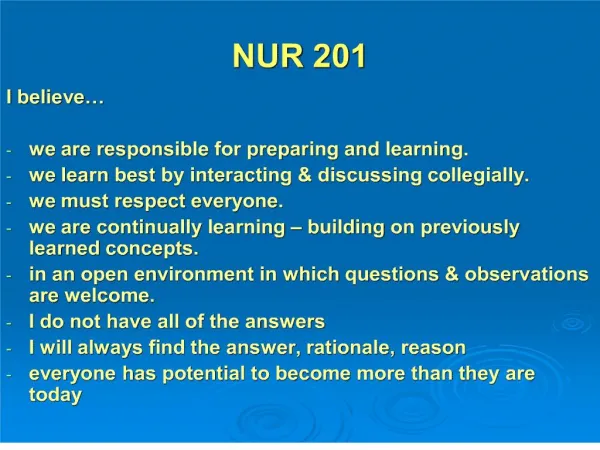
1. NUR 201 I believe�
we are responsible for preparing and learning.
we learn best by interacting & discussing collegially.
we must respect everyone.
we are continually learning � building on previously learned concepts.
in an open environment in which questions & observations are welcome.
I do not have all of the answers
I will always find the answer, rationale, reason
everyone has potential to become more than they are today
3. Content Approach Anatomy & Physiology Review
Demographics/occurrence
Pathophysiology
Clinical Picture
Medical Management
Nursing Process (APIE)
Assessment - Nursing Actions - Education
4. Interferences with VentilationRespiratory Anatomy & Physiology Anatomy
Structure of the Chest Wall: Ribs, pleura, muscles of respiration
Upper Respiratory: nose, pharynx, adenoids, tonsils, epiglottis, larynx, and trachea
Lower Respiratory: bronchi, bronchioles, alveolar ducts, and alveoli
Physiology
Ventilation: inspiration and expiration
Elastic Recoil: elastin fibers that recoil after expansion
Diffusion: Exchange of oxygen and carbon dioxide
Arterial Blood Gases / Oximetry
5. Lungs A & P Review
6. Respiratory AssessmentA&P Review
7. Thorax Anatomical Landmarks
8. Interferences with VentilationAlveolar Gas Exchange
9. Interferences with VentilationAssessment History
Cues to Respiratory Problems:
Shortness of breath � dyspnea
Orthopnea / Nocturnal dyspnea
Wheezing
Cough / sputum production
Hemoptysis
Voice change
Fatigue
10. Interferences with Ventilation Assessment Thorax & Lungs
Inspection:
Posture, chest movement, abnormalities of sternum
Respiratory rate, depth, rhythm
Palpation:
Equality of chest expansion
Tactile Fremitus
Percussion:
Hyperresonance
Dullness
Auscultation:
Discontinuous: fine crackles/rales / coarse crackles / rales
Continuous: Wheeze, Rhonchi
Pleural friction rub
11. Respiratory AssessmentPercussion
12. Larynx Anatomical Landmarks
13. Respiratory Assessment
14. Respiratory AssessmentAscultation Landmarks
15. Respiratory AssessmentBreath Sounds
16. Respiratory AssessmentNormal Breath Sounds
17. Respiratory AssessmentAdventitious Breath Sounds
19. Interferences with VentilationDiagnostic Studies
Blood Studies: Hgb, Hct, ABGs
Sputum Studies: C&S, Gram Stain, Acid-fast smear; Cytology
Radiology:
Chest x-ray-- posterior-anterior / lateral
Computed tomography (CT) � cross sections of the lung with and without contrast � used often
Magnetic resonance imaging (MRI) � images of pulmonary structures � limited use
Pulmonary angiogram � x-rays after injection of radiopaque dye� used to dx pulmonary embolism
Positron emission tomography (PET) � IV glucose administration � malignant tumors show increased uptake of glucose
Ventilation-Perfusion Scan � Perfusion: isotope administration which outlines pulmonary vasculature; Vent: inhalation of radioactive gas which outlines the alveoli � dx pulmonary emboli
20. Interferences with VentilationDiagnostic Studies Endoscopic Exams (done in x-ray or OR):
Bronchoscopy � fiberoptic visualization of bronchi � biopsy; also used to remove mucous plugs, foreign bodies, obstructions
Mediastinoscopy � scope through a small incision n the suprasternal notch � visualize mediastinum for tumors, lymph nodes, infections, sarcoidosis
Biopsy: Transbronchial or open lung biopsy � done in x-ray or OR
Thoracentesis � insertion of a needle into the pleural space � pleural fluid, install medication - done at bedside
Pulmonary Function Testing � tests to measure lung volumes and used to dx pulmonary disease, monitor progress, evaluate disability, evaluate response to bronchodilators � done in pulmonary lab
Skin Testing � intradermal planning of test dose to assess skin reaction by measuring mm induration � TB, various lung diseases
21. Pulmonary Function Test Relationship of Lung Volumes & Capacities
22. Respiratory Diagnostic Testing Fiberoptic Bronchoscopy
23. Diagnostic Lung Tests Thoracentesis
24. Pair Share � Critical Thinking Upon performing a lung sound assessment of the anterior chest, the nurse hears moderately loud sounds on inspiration that are equal in length with expiration. Where in the airway would this lung sound be considered normal?
a. Trachea
b. Primary bronchi
c. Lung fields
d. Larynx
25. Pair Share � Critical Thinking The name that describes the particular lung sound in the previous questions is which of the following?
a. Bronchial
b. Bronchovesicular
c. Vesicular
d. Basilar
26. Interferences with VentilationRegulation of Acid-Base BalanceReview
Acid � contributes hydrogen ion
Two types:
Volatile respiratory acid
Dehydrates and excreted in the form of a gas
Nonvolatile metabolic acid
Metabolized and excreted in the form of body fluids
27. Interferences with VentilationRegulation of Acid-Base Balance Review Base � accepts or removes hydrogen ion
Buffer- controls the hydrogen ion concentration:
Absorbing hydrogen ions when an acid is added OR
Releasing hydrogen ions when base is added.
Three Buffer Systems:
Bicarbonate � operates in lungs & kidneys
Phosphate � renal tubules
Protein � Hgb, plasma proteins, & intracellular protein
28. Interferences with VentilationRegulation of Acid-Base Balance Factors to remember:
Lungs � Eliminate or retain carbon dioxide C02
Kidneys � excrete or form bicarbonate HC03
Food � converted by the body � H20 + CO2 + energy
Lung Kidney
C02 + H20 = H2CO3 = HCO3- + H+
29. Interferences with VentilationNormal Acid-Base Balance
30. Interferences with VentilationRegulation of Acid-Base Balance Lungs/Respiratory System
Increase or decrease hydrogen ion concentration
Through respiratory rate and depth
Result: C02 is either retained or eliminated
Changes can occur within minutes
Controlled in the medulla oblongata�respiratory center
> = increased; < = decreased
<pH causes > respirations = <C02 + correcting pH
>pH causes < respirations = >C02 + correcting pH
31. Interferences with VentilationRegulation of Acid-Base Balance Renal System
Reabsorb and conserve bicarbonate
Can generate additional bicarbonate and eliminate excess hydrogen ions as compensation for acidosis
Three mechanisms:
Secretion of small amounts of free hydrogen into the renal tubule
Combination of hydrogen ions with ammonium
to form ammonium
Excretion of weak acids
Urine pH 4 � 8
32. Interferences with VentilationRegulation of Acid-Base Balance> = increased; < = decreased
33. Respiratory Respiratory Alkalosis Acidosis
34. Acid-Base ImbalanceRespiratory Acidosis Hypoventilation from primary lung problem
Atelectasis
Pneumonia
Respiratory failure
Airway obstruction
Chest wall injury
Cystic fibrosis
Hypoventilation from other factors
Drug overdose
Head injury
Paralysis of respiratory muscles
Obesity
35. Acid-Base ImbalanceRespiratory Alkalosis Hyperventilation from primary lung problem
Asthma
Pneumonia
Inappropriate ventilator settings
Hyperventilation from other factors
Anxiety
Disorders of the central nervous system
Salicylate overdose
36. Interferences with Ventilation Regulation of Acid-Base Balance Respiratory Function
37. Pair Share � Critical Thinking
What acid-base imbalance would you suspect for the patient having respiratory problems with respiratory rate: 28/min and expiratory wheezing?
38. Pair Share � Critical Thinking
What acid-base imbalance would you suspect for the post-operative patient with respiratory rate 10/min, difficulty to arouse, but arouses with verbal stimuli
39. Interferences with VentilationRegulation of Acid-Base Balance> = increased; < = decreased
40. Metabolic MetabolicAlkalosis Acidosis
41. Acid-Base ImbalanceMetabolic Acidosis
Starvation
Diabetic ketoacidosis
Renal failure
Lactic acidosis from heavy exercise
Use of drugs (ASA, methanol, ethanol)
Acute renal tubular necrosis
Diarrhea
42. Acid-Base ImbalanceMetabolic Alkalosis Excessive vomiting
Prolonged nasogastric suctioning
Hypokalemia or hypocalcemia
Excess aldosterone
Use of drugs (steroids, sodium bicarbonate, diuretics)
43. Interferences with Ventilation Regulation of Acid-Base BalanceMetabolic Function
44. Interferences with Ventilation Regulation of Acid-Base Balance
Normal Values:
45. Interferences with Ventilation Regulation of Acid-Base BalanceArterial Blood Gas Intrepretation > = increased; < = decreased Step 1: Evaluate the pH
pH <7.35 = acidosis
pH >7.45 = alkalosis
Step 2: Evaluate Respiratory Function
Paco2 >45 mm HG = ventilatory failure & respiratory acidosis
Paco2 <35 mm HG = hyperventilation & respiratory alkalosis
46.
Step 3: Evaluate Metabolic Processes
Serum bicarbonate HCO3 <22 mEq/L = metabolic acidosis
Serum bicarbonate HCO3 >26 mEq/L = metabolic alkalosis
Step 4: Determine the Primary Disorder
When Paco2 & HCO3 are both abnormal:
Determine which follows the deviation from the pH
and
Deviates the most from normal
48.
Respiratory Acidosis:
49.
Respiratory Alkalosis:
50. Respiratory AssessmentRelationship between PaO2 & SpO2
51. Respiratory AssessmentPulse Oximetry
52. Metabolic Acidosis:
53. Metabolic Alkalosis
54. Pair Share � Critical Thinking Arterial Blood Gas nterpretation
Interpret the following ABGs: pH - 7.50
Paco2 � 28; HCO3- - 25; Pao2 - 88
A. Acute exacerbation of asthma.
B. Renal failure.
C. Acute exacerbation of COPD.
56. Pair Share � Critical Thinking Arterial Blood Gas Interpretation Interpret the following ABGs: pH -7.28;
Paco2 � 52; HCO3- - 26; Pao2 - 68
A. Acute exacerbation of asthma.
B. Diabetic ketoacidosis.
C. Acute exacerbation of emphysema
D. Patient with prolonged NG drainage
57. Pair Share � Critical Thinking Arterial Blood Gas Interpretation
Medical Dx: Acute exacerbation of emphysema
58. Pair Share � Critical Thinking Arterial Blood Gas Interpretation
Medical Dx: Acute exacerbation of emphysema
59. Pair Share � Critical Thinking Arterial Blood Gas Interpretation Interpret the following ABGs:
pH - 7.30; Paco2 � 37; HCO3- - 18 ; Pao2 - 90
A. Acute exacerbation of emphysema.
B. Acute episode of asthma.
C. Renal failure.
60. Pair Share � Critical Thinking Arterial Blood Gas Interpretation
Medical Dx: Renal Failure
61. Interpret the following ABGs:
pH � 7.48; Paco2 � 45;
HCO3- - 32; Pao2 � 98
I
Acute renal failure.
Acute diabetic ketoacidosis.
Patient with prolonged NG drainage.
Patient with prolonged diarrhea.
62. Pair Share � Critical Thinking Arterial Blood Gas Interpretation
Medical Dx: postop patient with NG with large amount of NG output
63. Interferences with VentilationClassification of Resp Failure Hypoxemic PaO2 <= 60 mmHg on 60% O2
Acute Respiratory Distress Syndrome
Direct lung injury: aspiration; severe, disseminated pulmonary infection; near-drowning; toxic gas inhalation; airway contusion
Indirect lung injury: sepsis/septic shock; severe
non-thoracic trauma, cardiopulmonary bypass
Pathophysiology �
Fluid enters interstitial space and alveoli�impaired gas exchange
< PaO2 and > PaCO2.
Ischemia to pulmonary capillaries
< integrity of the alveolar-capillary membrane
64. Interferences with VentilationResp Failure � Medical Tx Goals Maintain adequate oxygenation & ventilation
Oxygen therapy
Mobilization of secretions
Effective coughing and positioning
Hydration & humidification
Chest physical therapy
Airway suctioning
Positive pressure ventilation
Relief of bronchospasm
Reduction of airway inflammation
Reduction of pulmonary congestion
Treatment of pulmonary infections
Reduction of severe anxiety, pain, and agitation
Treat underlying cause
Maintain adequate cardiac output
Maintain adequate hemoglobin concentration
65. Interferences with VentilationNursing Diagnosis
66. Interferences with VentilationNursing Diagnosis Ineffective airway clearance
Ineffective breathing pattern
Risk for imbalanced fluid volume
Anxiety
Impaired gas exchange
Imbalanced nutrition: less than body requirements
67. Interferences with VentilationClassification of Resp Failure Hypercapnic PaCO2 > 45 and pH < 7.35
Imbalance between ventilatory supply and ventilatory demand
Supply: maximum ventilation that the pt. can sustain without developing respiratory muscle fatigue
Demand: The amount of ventilatory needed to keep the PaCO2 within normal limits
Normally: supply > demand
Hypercapnia � ventilatory failure � inability of the respiratory system to ventilate out sufficient CO2 to maintain a normal PaCO2
68. Interferences with VentilationRespiratory Failure Causes of hypercapnic respiratory failure
Airways & alveoli
Asthma, emphysema, chronic bronchitis, cystic fibrosis
Central nervous system
Problems that suppress the drive to breathe � drug overdose, brainstem infarction, severe head injury, spinal cord injuries
Chest wall
Flail chest, fractures, kyphoscoliosis, massive obesity
Neuromuscular conditions
Guillain-Barre syndrome, muscular dystrophy, multiple sclerosis, myasthenia gravis, ALS
69. Interferences with VentilationNursing Management Tracheostomy Care Indications for Tracheostomy
Bypass an upper airway obstruction
Cases of prolonged intubation & mechanical ventilation
Facilitate removal of secretions
Permit oral intake & speech in a patient who requires long-term mechanical ventilation
70. Interferences with VentilationNursing Management Tracheostomy Care Types of Tracheostomy Tubes
Shiley & Portex fenestrated tracheostomy tube with cuff, inner cannula, decannulation plugs & pilot balloon
Fenestrated: openings on the surface of the outer cannula that permit air from the lungs to flow over the vocal cords
Allows the patient to breathe spontaneously, speak, & cough up secretions
Used by the patient who can swallow without risk of aspiration but requires suctioning for secretion removal.
Used by the patient who requires mechanical ventilation for fewer than 24 hours a day
Bivona (Fome) tracheostomy tube with foam cuff and obturator
71. Interferences with VentilationNursing Management Tracheostomy Care Inserting trach tube
Removing obturator
Trach tube maintenance
Cuff � deflated versus inflated
Trach suctioning
Sterile procedures
Use only Normal sterile saline (NO Water)
Apply suction as catheter is withdrawn
Cleaning procedure
Disposable inner cannula
Skin care; Changing trach ties
72. Interferences with VentilationNursing Management Patient with a ET Tube or Trach Maintain correct tube placement
Breath sounds
Maintain proper cuff inflation
Cuff pressure 20-25 mm Hg (capillary perfusion 30 mm Hg)
Monitor Oxygenation & Ventilation
ABGs; pulse oximetry;
PETCO2 (partial pressure of end-tidal CO2
Respiratory rate assessment
73. Interferences with VentilationNursing Management Patient with a ET Tube or Trach Maintain tube patency
Closed and open suctioning
Assess patient:
Visible secretions
Sudden onset of respiratory distress
Suspected aspiration
Increase in peak airway pressure
Auscultation of adventitious breath sounds
Increased respiratory rate / increased cough
Sudden or gradual decrease in Pao2 and/or SpO2
74. Interferences with VentilationNursing Management Patient with a ET Tube or Trach Providing Oral Care and Maintaining Skin Integrity
Lips, mouth, teeth, tongue, oropharynx q2hr
Prevent pressure from ET tube
Ensure ET or Trach is secured properly
Change securing tapes qd & prn
Fostering Comfort and Communication
Position of comfort
Include family in care as appropriate
Pain relief
Methods of communication: pictures, word chart
75. Interferences with VentilationNursing Management Patient with a Trach Nursing Diagnoses:
Ineffective airway clearance
Impaired verbal communication
Risk for infection
Imbalanced nutrition
Impaired swallowing
Ineffective therapeutic regimen management
Potential complication�hypoxemia related to misplaced or properly functioning trach tube
76. Tracheostomy TubeVelcro Collar
77. Anatomy of the Tracheostomy Tube
78. Fenestrated Tracheostomy Tube
79. Tracheostomy Suctioning
80. Pair Share � Critical Thinking Humidification of the air is essential for the patient with an artificial airway because humidification helps do which of the following?
1. Prevent tracheal damage
2. Promote thick secretions
3. Dry out the airways
4. Liquefy the secretions
81. Interferences with VentilationNursing ManagementCare of the Patient Requiring Mechanical Ventilation Types of mechanical ventilation
Settings
Modes of volume ventilation
Complications
Nutritional therapy
Weaning from positive pressure ventilation & extubation
Home mechanical ventilation
82. Interferences with VentilationNursing ManagementCare of the Patient Requiring Mechanical Ventilation
Types of mechanical ventilation
Controlled mandatory ventilation (CMV) � breaths are delivered at a set rate and volume independent of the patient�s respirations
Assist-control mechanical ventilation � delivered preset volume & frequency; when pt. initiates a spontaneous breath, a full volume is delivered
Synchronized intermittent mandatory ventilation � prevent volume at a frequency synchronized with patient�s respirations � most common form used
83. Interferences with VentilationNursing ManagementCare of the Patient Requiring Mechanical Ventilation Other Ventilatory Maneuvers:
Positive end-expiratory pressure (PEEP) -- + pressure applied during expiration � increases functional residual capacity � prevents alveoli collapse
Continuous positive airway pressure (CPAP) � prevents patients airway pressure from falling to zero � Used for sleep apnea
High-frequency ventilation � delivery of small tidal volume at a rapid respiratory rate � maintains lung volume and reduces intrapulmonary shunting
84. Interferences with VentilationNursing ManagementComplications of Mechanical Vent Pulmonary
Barotrauma
Volu - pressure trauma
Alveolar hypoventilation
Alveolar hyperventilation
Ventilator-assisted pneumonia
Sodium & Water Imbalance � Na+ & fluid retention
Neurologic � impaired cerebral blood floor
Gastrointestinal � stress ulcer/GI bleed
Musculoskeletal � contractures, pressure ulcers, footdrop, complications of immobility
Psychosocial = pain, fear, isolation, anxiety, dependency
Mechanical Disconnnection or Malfunction � Alarm Alert
85. Establish a nutritional program if the pt is to be without food for 3-5 days
Total parenteral nutrition
Enteral feedings
Concern:
Aspiration
Increased carbohydrates = increases CO2 levels
86. Interferences with VentilationNursing ManagementWeaning the Pt. from Mechanical Ventilation & Extubation Weaning: reducing ventilator support & resuming spontaneous ventilation
Pre-weaning � assessing Pt respiratory effort
Muscle strength; PEEP; TV; VC; clear lungs
Weaning � SIMV � synchronized intermittent mandatory ventilation; CPAP; humidified T-piece
Outcome Phase � extubation after hyperoxygenation & suctioning prior to removal; supplemental oxygen, monitoring, need for re-intubation
87. Interferences with VentilationNursing ManagementHome Mechanical Ventilation Negative & positive pressure ventilators
Settings and alarms
Home Health / Family participation in care
Decreased risk for nosocomial infection
Concerns:
Reimbursement: home health, disposable products may be nonreimbursable
Family � respite care
88. Pair Share � Critical Thinking A 72-year old female brought to the ER following a head-on car accident. She has blunt injury to the chest�difficulty breathing, cyanosis; receiving O2 4l/min NC.
Assessment priority?
Immediate nursing actions