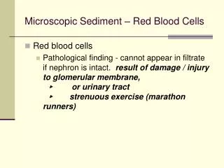
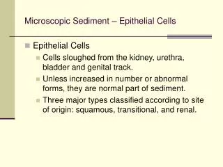
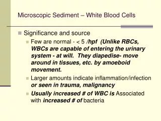
Microscopic Sediment – Miscellaneous
Microscopic Sediment – Miscellaneous. Miscellaneous urine sediment structures Mucous - threadlike, transparent. Low light is needed in order to be able to see mucous threads. Usually a vaginal contaminant. Do not confuse with casts. Microscopic Sediment – Miscellaneous.

Microscopic Sediment – Miscellaneous
E N D
Presentation Transcript
Microscopic Sediment – Miscellaneous • Miscellaneous urine sediment structures • Mucous - threadlike, transparent. Low light is needed in order to be able to see mucous threads. Usually a vaginal contaminant. Do not confuse with casts.
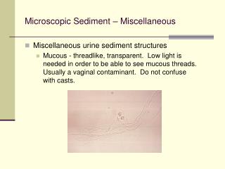
Microscopic Sediment – Miscellaneous • Mucous threads (2 wbc within, 1 rbc within, 1 rbc out) • 160x magnification
Microscopic Sediment – Miscellaneous • Mucous threads
Microscopic Sediment – Miscellaneous • Miscellaneous urine sediment structures • Bacteria – May be a contaminate from poor collection or transport. or as the result of spec sitting at RT too long. Make the urine alkaline. We quantitate but do not describe (ie rods, cocci, etc.) or try to speciate. E. coli is the most common cause of UTI. In UTI, usually see increased number of WBCs.
Microscopic Sediment – Miscellaneous • Miscellaneous urine sediment structures • Squamous epithelial cells and bacteria x250
Microscopic Sediment – Miscellaneous • Miscellaneous urine sediment structures • WBC and bacteria, S-M stain x 100
Microscopic Sediment – Miscellaneous • Miscellaneous urine sediment structures • Yeast – May be a contaminant since it is normal vaginal flora. But may be seen in diabetics (since increased glucose in urine), or a patient on antibiotic treatment which suppresses other flora allowing yeast to overgrow
Microscopic Sediment – Miscellaneous • Budding yeast with squamous epithelial cell • 200x (no segs, contamination? vs infection)
Microscopic Sediment – Miscellaneous • Miscellaneous urine sediment structures • Yeast with hyphae, interference contrast microscopy x 160
Microscopic Sediment – Miscellaneous • Miscellaneous urine sediment structures • Parasites – usually a vaginal or fecal contaminant • Trichomonas - resembles a WBC but has a flagellum and moves. Can be found in males or females. A sexually transmitted parasite, can also be picked up in nature.
Microscopic Sediment – Miscellaneous • Miscellaneous urine sediment structures • Fecal parasites - pinworm (E. vermicularis), other intestinal parasites. • Urine - bladder parasite Schistosoma haematobium
Microscopic Sediment – Miscellaneous • Miscellaneous urine sediment structures • Sperm - whether or not to report in a UA depends on your lab. If in doubt, report. Can indicate prostatic problems. in older men. • in ♀ a vaginal contaminant, tells dr that pt is sexually active.
Microscopic Sediment – Miscellaneous • Spermatozoa
Microscopic Sediment – Miscellaneous • Miscellaneous urine sediment structures • Oval fat bodies - degenerating or necrotic renal epithelial cells /possibly macrophages contain fat globules (bi-refringent fat droplets) in their cytoplasm. • Rare, but serious - seen in nephrotic syndrome. If fat is cholesterol will polarize (oval fat bodies, fat casts, uric acid , starch crystals - all sorts of things).
Microscopic Sediment – Miscellaneous • Oval Fat Bodies • They are often (but not always) seen in specimens with increased protein (proteinuria) & nephrotic syndrome. • Source – fatty degeneration within renal epithelial cell cytoplasm, or possibly macrophages which contain fat (foam cells). (Authors vary, very possibly both exist.) • True oval fat bodies demonstrate maltese cross (due to presence of esterified cholesterol in liquid-crystal form) under polarized light.
Microscopic Sediment – Miscellaneous • Oval Fat Bodies cont. • Fat stain may assist in detection, but degenerated cells may also pick up stain. • Lipiduria • Lipid droplets seen as • Free bi-refringent fat droplets of various sizes • Intracellular fat in ‘oval fat bodies’ • Within the matrix of the ‘fatty cast’
Microscopic Sediment – Miscellaneous • Oval fat bodies • Hpf, bright field and polarized
Microscopic Sediment – Miscellaneous • Polarized oval fat bodies (maltese cross)
Microscopic Sediment – Miscellaneous • Case study: lymphoma pt. just started on chemo. pH 8, pos nitrate, pos leu esterase, trace protein, 80 wbc/hpf, 20 rbc/hpf.
Microscopic Sediment – Miscellaneous • Case study: lymphoma pt. just started on chemo. pH 8, pos nitrate, pos leu esterase, trace protein, 80 wbc/hpf, 20 rbc/hpf. • My thoughts: RTE with neutral fat or trig (since they do not polarize). Suggest fat stain.
Microscopic Sediment – Miscellaneous Artifacts • Artifacts - powder crystals, fibers, oils, hairs, pollen • Cotton fibers
Microscopic Sediment – Miscellaneous Artifacts • Starch granules
Microscopic Sediment – Miscellaneous Artifacts • Air bubbles